
Complications of Venous Leg Ulcers
Practice Accelerator
January 31, 2019
January 31, 2019
Keywords
Categories
Venous leg ulcers (VLUs) are difficult to treat, and when they are present a variety of complications may arise. These complications can be challenging to treat and may often contribute to the prolonged healing times resulting from chronicity found with many VLUs. Further, if the condition of the ulcer deteriorates, it may worsen any complication already present or serve as the catalyst for the development of complications.
Infection of Venous Leg Ulcers
The most common complication of venous leg ulcers is infection, which can arise from a variety of sources, including the following:1
- Bacterial: furuncles, ecthyma, mycobacterioses, syphilis, erysipelas, anthrax, diphtheria, chronic vegetative pyoderma, tropical ulcer
- Viral: herpes, variola virus infection, cytomegalovirus infection
- Fungal: sporotrichosis, histoplasmosis, blastomycosis, coccidioidomycosis
- Protozoal: leishmaniasis
When infection is present in the VLU, the patient may present with fever, wound fluid rich in leukocytes, increasing pain, cellulitis, necrotic tissue, and purulent exudate with or without odor. When signs of infection are present, the patient should be treated with appropriate systemic antibiotics, topical antimicrobials, or antiseptics.2 However, VLUs that are critically colonized with bacteria or bacterial biofilms without signs of systemic infection may be treated in multiple ways, including topical antibiotics. Sequential, aggressive debridement can also be used to treat infected ulcers.1
Venous Leg Ulcer Complications
Osteomyelitis and Septicemia
If infection is left unchecked, there is a risk that it will progress to osteomyelitis or even septicemia, which generally require intravenous antibiotics. Extreme cases may require amputation.
Cellulitis
Cellulitis is inflammation and infection of the skin, subcutaneous tissue, and lymphatic fluid of the skin1 that causes the area around it to be red, warm, painful, and tender to the touch. It is caused by a type of bacterial infection, generally streptococcal or staphylococcal, but it may be caused by other bacteria as well. It is generally a well-demarcated area (unlike venous eczema). It may cause deterioration of the ulcer and is typically treated with oral antibiotics.3
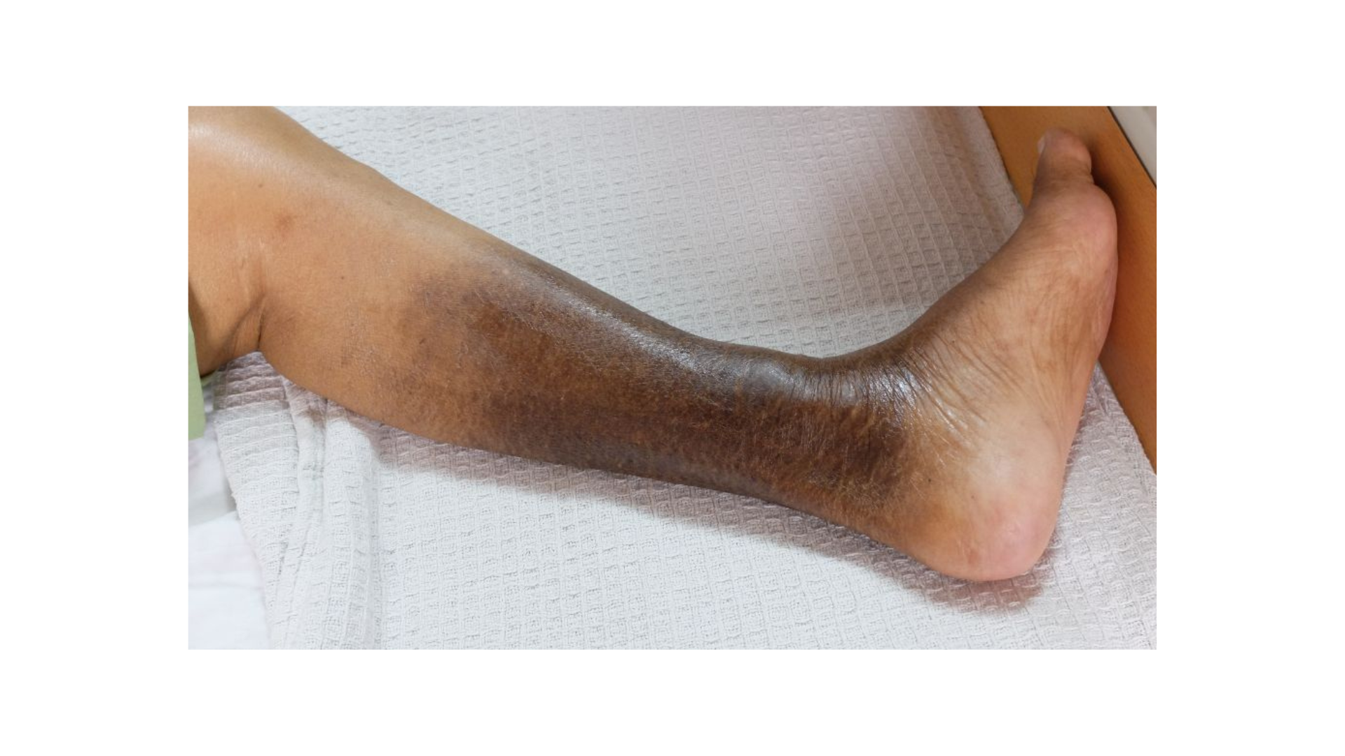
Venous Eczema
Venous eczema can be a precursor to an ulcer, and it can also persist in varying degrees of severity while an ulcer is present. Eczema found in a VLU can be gravitational eczema, stasis eczema, varicose eczema, or venous eczema, depending on its relationship with the underlying cause. It can also be acute or chronic.4 Mild eczema will be itchy, red and swollen, and scaly or flaky. In more severe cases, the skin may discolor to brown or red and become tender and tight, and small white scars called atrophie blanche may develop.5 Hyperkeratosis may occur when the scales from the eczema become thickened. Relief may be found when an emollient is applied to the affected area.3 Additional treatments can include a moderate to highly potent corticosteroid, antiseptic, and astringent, as well as oral antibiotics if the eczema becomes infected.3
Malignancy
Malignancy of ulcers is rare, although any VLU with atypical features should be examined by biopsy. For those ulcers that do not close within four to six weeks with standard compression therapy and wound care, a biopsy can provide valuable information.1 Malignancy tends to happen when treatment of the ulcer is delayed, and the ulcer persists for a prolonged period of time. The majority of malignancies found in ulcers are squamous cell carcinomas. Patients with malignancy of an ulcer may develop worsening pain, bleeding, or tissue necrosis. 6
Conclusion
Characteristically, differences in VLUs can help identify and differentiate diagnosis for more precise treatment. VLUs tend to be chronic, lasting weeks, months, and sometimes years. Complications can include cellulitis and other infections including osteomyelitis, malignancy, and even amputation and death. Management of bioburden, infection, and skin conditions is crucial in moving toward healing and preventing recurrence.
References
1. O'Donnell TF Jr, Passman MA, Marston WA, et al. Management of venous leg ulcers: clinical practice guidelines of the Society for Vascular Surgery and the American Venous Forum. J Vasc Surg. 2014;60(2):3S–59S.
2. O'Meara S, Al-Kurdi D, Ologun Y, et al. Antibiotics and antiseptics for venous leg ulcers. Cochrane Database Syst. Rev. 2014;(1):CD003557.
3. Grey JE, Harding KG. Venous and arterial leg ulcers. BMJ. 2006;332(7537):347–50.
4. Newton H. Eczema associated with venous leg ulcers. Wound Essentials. 2014;9(2):72–8.
5. UK National Health Service. Overview – varicose eczema. London, United Kingdom: National Health Service; 2016 . https://www.nhs.uk/conditions/varicose-eczema/. Accessed January 7, 2019.
6. Smith J, Mello LB, Nogueira Neto NC, et al. Malignancy in chronic ulcers and scars of the leg (Marjolin's ulcer): a study of 21 patients. Skeletal Radiol. 2001;30(6):331–7.
The views and opinions expressed in this blog are solely those of the author, and do not represent the views of WoundSource, HMP Global, its affiliates, or subsidiary companies.


Follow WoundSource
Tweets by WoundSource