Creepy Crawlies of Wound Care
October 15, 2020
As summer begins to wind down and we look ahead to Halloween, let’s discuss some “creepy crawlies” we may encounter in wound care that may cause apprehension in even the most seasoned health care staff.
Scabies
This highly contagious, itchy rash may manifest when the skin is infected with Sarcoptes scabiei, a mite that is transmitted between individuals via direct skin contact and, less commonly, from fomites such as clothing and linen.1 The mites burrow into the skin, where their presence (and their feces) triggers an immune reaction from the host. The most common symptoms include pruritus that is worse at night, thin burrow marks, and a linear maculopapular rash that often manifests on finger and toe web spaces, armpits, and the groin.
Treatment typically involves applying permethrin 5% cream over the entire body from the neck down, letting it sit for 8 to 14 hours, and repeating this application seven days later; a resistant or “crusted” infestation may involve use of an oral antiparasitic.1 It is very important to treat all household members and close contacts, as well as properly launder all linens and clothing in hot water and a high- heat dryer to reduce the risk of re-infestation.
Maggots
Sterile maggots are sometimes used in wound debridement, but feral (wild) maggots are contaminants that can cause and spread infection and should be removed. There are actually several types of myiasis (infestation of vertebrates with fly larvae), which are classified by the anatomical area of invasion.2 Although myiasis can occur in cavities such as the eye, ears, nose, and genitals, the most common type is cutaneous (skin) myiasis. Most commonly transmitted in warm and wet climates, furuncular myiasis occurs when a larva (most commonly a botfly larva ) penetrates the skin and causes a furuncle to form containing one or more maggots, depending on the species.2 This “boil” is erythematous and painful at night, and may it drain serosanguinous or purulent exudate.
Many patients have the sensation of movement in the lesion, especially preceding the eruption of drainage. Treatment involves removing the larva via inducing hypoxia by smothering the maggot(s) with an occlusive dressing or substance (petrolatum) to force the offender to emerge, or surgical removal.2 Migratory myiasis involves burrowing of larva into the epidermis, but it is usually self-limiting because these maggots are unable to complete their life cycle in human skin. Wound myiasis occurs when a fly lays eggs in a wound, often attracted by the odor of necrotic or purulent wounds, especially alkaline exudate. Wound patients with poor hygiene, low socioeconomic status, impaired vision, and restricted mobility are at risk, but even wounds that are properly cared for or dressed can develop myiasis because of the penetration of larva through non-occlusive dressings.
Treatment necessitates removal of all larvae, although there is no consensus on the best method for this. Some recommend irrigation with isopropyl alcohol, hydrogen peroxide, iodine, or hypochlorite solutions, but those solutions can be cytotoxic to the wound, and they are not proven to kill all of the maggots successfully.3 Removing the maggots by manual means may be safer to the wound bed as well as more cost effective. Suffocating the maggots with a thick layer of petrolatum may encourage migration from holes and cavities to facilitate manual removal with gauze or forceps; Elder and Grover recommend using a Yankauer suction device to remove and contain the maggots.4
Leeches
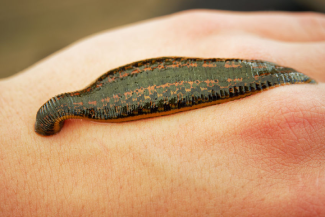
Leeches, like maggots, can be both friends and foes from a medical standpoint. Wild leeches can be either aquatic or terrestrial, and they ingest the blood of humans and mammals through the skin or internal mucous membranes. The saliva of a leech contains antithrombins, vasodilators, and platelet aggregation inhibitors that promote bleeding, which on average lasts for 10 hours after the leech is removed.5 Rarely, an individual with multiple leech bites could have anemia from the acute blood loss. Removal of the leech can be done with saline solution, which causes the leech to release. Use caution when manually removing leeches because squeezing the organism can eject its contents into the wound, thus risking infection and even blood-borne infectious diseases.5
Ensure that the entire leech is removed because remaining parts left in the skin will prolong bleeding and infection risk. Control bleeding from the wound with a hemostatic agent or bandage and/or a pressure dressing. The blood-sucking creatures may be scary in the wild, but they also are used for medicinal purposes. Medicinal leech therapy (MLT) can be applied to improve circulation in surgical flaps or skin grafts, reduce pain and inflammation, and to act as an anticoagulant.6 MLT can assist in improving blood flow after microsurgery or after revascularization, it is also used in the treatment and prevention of venous congestion.7 Bacteriostatic effects have also been noted with Staphylococcus aureus, Pseudomonas aeruginosa, and Escherichia coli. Risks of MLT include anemia, infection with Aeromonas bacteria from the leech, lymphadenopathy, and scarring at the application site.6
Conclusion
As squeamish as these parasites may make the caregivers tasked with their removal, we can also appreciate how stressful it can be for a patient, even if these maggots or leeches are placed as part of a medical treatment. If a patient presents with scabies, maggot infestation, or wild leech bites, it is important to check your institution’s infection control policies to appropriately contain, treat, and disinfect properly. Infection control is a concern with any parasite, so even after removal it is important to monitor the patient for signs of infection; providers may consider prophylactic antibiotics at their discretion or to treat a secondary infection.
Regardless of whether these “creepy crawlies” are placed for therapeutic benefit or occur from feral infestation, it is important to treat our patients with dignity and respect and to ease the anxieties of our patients afflicted with these unpleasant infestations.
References
1. Tarbox M, Walker K, Tan M. Scabies. JAMA. 2018;320(6):612. doi:10.1001/jama.2018.7480
2. Francesconi F, Lupi O. Myiasis. Clin Microbiol Rev. 2012;25(1):79-105.
3. McIntosh MD, Merritt RW, Kolar RE, Kimbirauskas RK. Effectiveness of wound cleansing treatments on maggot (Diptera, Calliphoridae) mortality. Forensic Sci Int. 2011;210(1-3):12-15. doi:10.1016/j.forsciint.2011.01.028
4. Elder J, Grover C. Wound debridement: lessons learned of when and how to remove “wild” maggots. J Emerg Med. 2013;45(4):585-587.
5. Conley K, Jamal Z, Juergens AL. Leech bite. In: StatPearls [Internet]. Treasure Island, FL: StatPearls Publishing; 2020. https://www.ncbi.nlm.nih.gov/books/NBK518971/ Accessed September 27, 2020.
6. Sig A, Guney M, Uskudar Guclu A, Ozmen E. 2017. Medicinal leech therapy—an overall perspective. Integr Med Res. 2017;6(4):337-343.
7. Abdualkader AM, Ghawi AM, Alaama M, Awang M, Merzouk A. Leech therapeutic applications. Indian J Pharm Sci. 2013;75(2):127-137.
About the Author
Lauren graduated with a BSN from the University of Buffalo in Western New York, where she was born and raised. She has held various nursing jobs, but continued to work towards a goal of a career in wound care nursing after she was one of only two students who signed up for a wound care clinical during nursing school. She currently works at the Advanced Wound Healing Center in Orchard Park, NY where she once had her nursing clinicals. She became credentialed in Wound, Ostomy, and Continence Nursing in 2019 and is incorporating her knowledge and skills into her busy clinic practice. When not at work, Lauren enjoys indoor spinning, playing guitar, video games, and rooting for the Buffalo Bills NFL team.
The views and opinions expressed in this blog are solely those of the author, and do not represent the views of WoundSource, HMP Global, its affiliates, or subsidiary companies.


Follow WoundSource
Tweets by WoundSource