Hypergranulation Tissue: What It Is and How to Treat
May 5, 2023
Keywords
Categories
Introduction
The small bright red cobblestone texture of healthy granulation tissue is just that: a granule of new collagen and the new growth of capillaries. Hypergranulation (Figure 1) is the excessive growth of granulation tissue, a symptom of a dysfunctional wound environment. Providers can identify hypergranulation by its appearance. As opposed to the bright, bubbly red appearance of healthy granulation tissue, unhealthy granulation, and hypergranulation tissue can present as large lobes of red tissue, and the coloration can vary between bright red and darker shades. These lobes will typically sit above skin level.
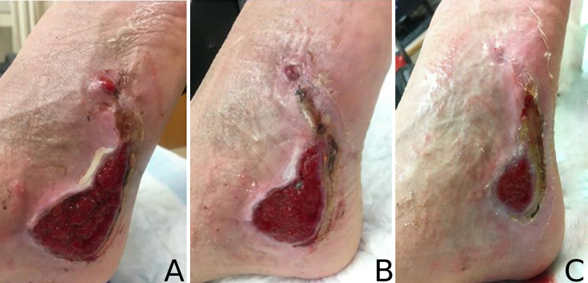 Figure 1: A. Hypergranulation to right lateral foot due to biofilm. B. Hypergranulation 1 week following of sharp debridement and rigorous wound hygiene. C. Resolution of hypergranulation at week 4 following continued sharp debridement, silver nitrate application, and rigorous wound hygiene.
This growth may be due to extrinsic factors, like excessive bacterial load and moisture, or intrinsic factors that can also contribute to excess inflammation and nutritional deficiencies.1
How Does Hypergranulation Tissue Stall Healing
Hypergranulation tissue tends to be friable, meaning it easily bleeds. When located next to a stoma, the serous and sanguineous drainage from hypergranulation tissue can contribute to poor pouch adherence and, ultimately, issues with leaking ostomy appliances. In hard-to-heal wounds, hypergranulation tissue can proliferate over the wound's edges, leading to early epithelial edge contact inhibition. This condition prevents further edge migration and can stagnate or completely stall wound healing. Addressing hypergranulation tissue should be coupled with managing any instigating factors: Excess moisture can be addressed with a more absorbent dressing, and inhibitory bacterial loads can be tampered down with non-cytotoxic antimicrobials. Several options address this over-proliferation of tissue, including debridement and some dressings.
Hypergranualtion Tissue Treatments
Debridement
Sharp, mechanical, and chemical debridement are all methods that can address hypergranulation. Each debridement method can be painful for patients if the hypergranulaton tissue is in a sensitive area. Patients are typically pre-medicated with a topical anesthetic such as lidocaine prior to debridement to address pain and allow for thorough debridement.
- Sharp debridement is a fast method of debridement that can also address several nonviable tissues, including the re-opening of epibolous edges. Limitations of sharp debridement include the licensing and training required to perform sharp debridement, bleeding, and pain.
- Chemical debridement uses the chemical silver nitrate.2 Silver nitrate cauterizes hypergranulation tissue, dampening down the over-proliferation of tissue with the added bonus of cauterizing the small blood vessels it encounters in doing so. Using silver nitrate to chemically cauterize hypergranulation tissue is a different procedure than using it for hemostasis following sharp debridement. The intent is to address hypergranulation tissue specifically.2 Despite pre-medication, patients can often experience a slight period of burning or stinging during or immediately following silver nitrate application. This discomfort is typically of short duration. Patients should be counseled that silver nitrate will turn the tissue a silver or black color and that this color will come off in their drainage. This tissue coloration is normal, and it should not cause alarm. Chemical cautery of hypergranulation tissue is also an accessible intervention that is frequently performed by certified wound and ostomy nurses.
- Mechanical debridement is accomplished with tools that include microfiber pads and cervical curettage wands. Mechanical debridement tends to be slower, and pain and bleeding can occur but are usually limited.
Topical Treatments
The application of a topical corticosteroid is another treatment option for hypergranulation tissue. Applying 0.1%-0.5% triamcinolone cream 2-3 times per day or triamcinolone ointment with less frequency can effectively treat hypergranulation tissue without the pain and stress associated with debridement.3 This method can be especially valuable in the pediatric population.4 Once the hypergranulation tissue is resolved, the steroid ointment should be stopped as atrophy and delayed wound healing can occur with continued use (Figure 2).
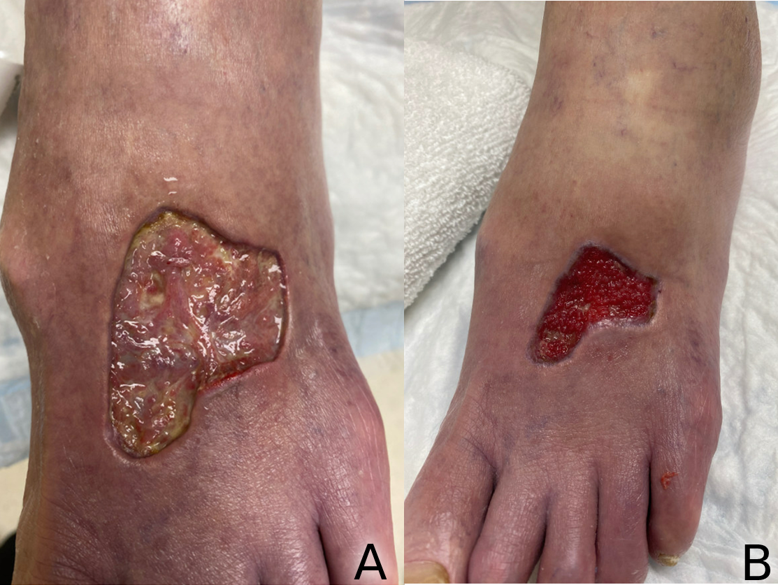 Figure 2: A. Left anterior foot after serial treatment with triamcinolone ointment. B. Left anterior foot 1 month following cessation of triamcinolone ointment. Note cobble-stoning of granulation tissue, and bright red healthy coloration.
Polyvinyl alcohol dressings (PVA) with antimicrobial pigments have been found to treat some cases of hypergranulation.1 Providers can use this method of hypergranulation topical treatment in combination with a topical corticosteroid. Using dressings as the sole treatment is most effective for mild hypergranulation. Large or deep lobes typically require additional treatment.
Conclusion
Hypergranulation tissue indicates a dysfunctional wound microenvironment, and it can lead to ostomy pouch leakage, delayed wound healing, or wound stagnation. Debridement, topical corticosteroid application, and PVA antimicrobial dressings are accessible treatment options. Due to the tendency for hypergranulation tissue to reform, serial treatments are often necessary until wound closure or the resolution of the peristomal hypergranulation.
References
- Mitchell A, Llumigusin D. The assessment and management of hypergranulation. Br J Nurs. 2021;30(5):S6-S10. doi:10.12968/bjon.2021.30.5.S6
- Ho C, Argaez C. Topical Silver Nitrate for the Management of Hemostasis: A Review of Clinical Effectiveness, Cost-Effectiveness, and Guidelines. Ottawa, Canada: Canadian Agency for Drugs and Technologies in Health; 2018. https://www.ncbi.nlm.nih.gov/books/NBK537873/.
- Bryant R, Nix D. Acute & Chronic Wounds: Current management concepts. 4th edition. St Louis, MO: Elsevier;2012.
- McShane DB, Bellet JS. Treatment of hypergranulation tissue with high potency topical corticosteroids in children. Pediatr Dermatol. 2012;29(5):675-678. doi:10.1111/j.1525-1470.2012.01724.x
About the Author
Laura Swoboda, DNP, APNP, FNP-BC, CWOCN-AP, is a Professor of Translational Science, Nurse Practitioner, and Wound Healing Coordinator at Froedtert & the Medical College of Wisconsin, where they advocate for nurse practitioners and nurse participation in research. They completed their Doctor of Nursing Practice degree at University of Wisconsin Milwaukee (UWM). Dr. Swoboda is a faculty member of the Clinical & Translational Science Institute of Southeast Wisconsin where they serve as principal investigator for quality improvement, evidence based practice, and research projects including the planning, implementation, management, and dissemination of projects in chronic wound care. They further participate in the research process in serving as a peer reviewer for scientific journals. Dr. Swoboda is on the National Pressure Injury Advisory Panel’s Prophylactic Dressing Standards Initiative Task Force, a member of the editorial board for the Wound Care Learning Network and Wound Management and Prevention, and on the board of directors for the WOCNCB and the AAWC.
The views and opinions expressed in this content are solely those of the contributor, and do not represent the views of WoundSource, HMP Global, its affiliates, or subsidiary companies.
More from this Author
// fixed missing link variable.
// fixed missing link variable.
// fixed missing link variable.