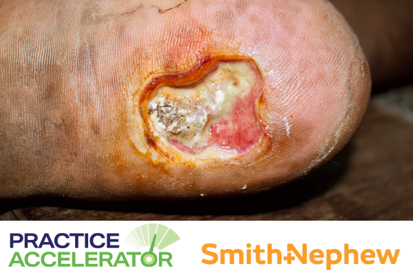
Biofilm: What is it?
Practice Accelerator
December 12, 2023
December 12, 2023
Keywords
Categories
When a wound becomes chronic after the clinician(s) has addressed underlying conditions, biofilm is often suspected. Biofilm complicates the detection and treatment of what otherwise might be manageable bacteria. This quick-forming, tenacious bacterial community is typically resistant to standard of care, thus requiring diligent, thorough strategies to undermine its detrimental hold on the wound healing cascade.1
What Is Biofilm?
Biofilm is a complex aggregate of potentially diverse bacterial communities.1 It is sessile, meaning the bacteria is fixed in place, rather than planktonic or free-floating.1 Biofilm impedes the patient’s immune response and prolongs inflammation. It prevents wound contracture, epithelialization, and other skin repair processes. Biofilm does not usually respond to standard wound management approaches. Biofilm and some of its bacterial components may be inherently antibiotic-resistant. Factors that further contribute to the hardiness of biofilm include2:
- High cell density
- Persister cells
- Nutrient and oxygen gradients
- Horizontal gene transfer
- Efflux activity
Persister cells are bacteria that can avoid the effects of antibiotics without changing their genetic makeup. Efflux activity is the movement of compounds that could disrupt the biofilm out of the cell.2
How Does Biofilm Develop?
Biofilm forms on the wound’s surface in 3 stages.3 First, bacterial cells attach and differentiate to form a structure, secreting a protective outer layer called the extracellular polymeric substance (EPS). The biofilm’s microcolonies grow under the protection of the EPS until the third stage when the fully mature biofilm structure is three-dimensional with small channels that facilitate nutrients, water, and waste as well as shelter microbes.3 Despite its complexities, this process can result in a mature biofilm within 24 hours.4 Redevelopment is more rapid, sometimes occurring 10 hours after sharp debridement, extending wound recidivism.1
What is an Extracellular Polymeric Substance (EPS)?
The EPS matrix thwarts antimicrobial access to the wound bed, making biofilms resistant to topical and systemic antibiotics.2 Biofilm may self-synthesize or develop this EPS from host-provided matrix matter attached to the wound’s surface, such as medical tubing or embed.1,3 Host substances that help form the EPS can include1,3:
- Proteins
- DNA
- Immunoglobulins
- Blood components
Research reports that these substances are 1,000-fold more tolerant to antimicrobial agents and disinfectants than planktonic cells. As such, this matrix protects the encased bacteria from the host immune response and antibiotics while fostering interaction among the encased bacterial species.1,3
Pathogenic Bacteria
Bacteria commonly found in biofilms may be categorized as commensal or pathogenic. Because they feature more upregulated genes, biofilms with pathogenic bacteria foster higher amounts of enzymes like matrix metalloproteinases. These enzymes facilitate the bacteria’s growth and movement, prolonging wound inflammation.3 Bacterial species commonly found in biofilm include2,3:
- Pseudomonas aeruginosa
- Staphylococcus aureus
- Enterococcus faecalis
- Coagulase-negative Staphylococci
- Proteus species
Generally, chronic wounds do not always exhibit clinical signs of infection, and diagnosing what bacteria species are present in biofilm may be difficult.1,5 Further, standard clinical microbiological (ie, culture) methods are typically unable to identify pathogens hidden in deep tissue or encased in biofilm.5 At present, point-of-care options for definitive biofilm detection are lacking.1
How Are Biofilms Detected?
Despite the high levels of suspicion often spurred by wound chronicity, bacteria hidden in biofilm are responsible for a high rate of false-negative cultures for wound infection, resulting in missed diagnoses.4 Identification of biofilm using the usual signs of infection, such as skin pH and temperature may not be reliable. Clinicians who aim to accurately diagnose the microbes present in biofilm may require the use of microbiological assays (culturing), molecular assays, and imaging.4 Microscopic techniques may be able to detect the presence of biofilm in tissue samples obtained during debridement, including4:
- Bright-field microscopy
- Scanning electron microscopy
- Confocal laser scanning microscopy
Several gene-related molecular techniques have been found to detect microbes in biofilm. A 2021 study contends that it may be possible to identify biofilm in wounds using a nitrocellulose membrane stained with a polysaccharide dye.5
How To Treat Biofilm: Debridement
Wound care professionals typically use wound debridement combined with antimicrobial therapy, when indicated, to treat biofilm.6
Several forms of debridement can be used, such as mechanical, chemical, or sharp debridement. Sharp debridement removes the biofilm and its niduses, but this removal could be incomplete. As previously stated, systemic antibiotics and topical antiseptics cannot typically overcome biofilm’s antibiotic resistance. Options like topical antiseptic application may forestall post-debridement biofilm spread and may be part of the recommendations for early therapy for stalled wounds.1 However, a review by Weigelt et al found that various topical products, such as honey and hypochlorous acid, didn't have enough evidence to support their use in biofilm management.1
How Much Do You Know About Biofilm and Infection? Take our quiz to find out! Click here.
Although silver used in vitro and in vivo has shown antimicrobial efficacy when used alone or incorporated into dressings, outcomes regarding biofilm eradication show little efficacy. Results of studies to show whether hyperbaric oxygen therapy can destroy biofilms are inconclusive.1 Bacteriophages (viruses) may be effective in eradicating the biofilm as well as the bacteria embedded within it.7 Ultrasound combined with antibiotic treatment, hydrogels, and products comprised of various types of nanoparticles and nanoemulsions have also been discussed as potential options to address biofilm.4
A category of products called electroceuticals has also appeared in the literature in the biofilm arena. Wireless bioelectric dressings used to treat biofilm utilize a direct current activated by wound exudate.1
Conclusion
Biofilm can harbor multiple species of dynamic and interactive microbes encased within a protective substance. The challenges of treating biofilm are numerous but not insurmountable. Researchers continue to explore ways to break down the outer barrier using both established (debridement) and novel (electroceutical and nanotechnology) approaches to disrupt biofilm’s microbial playing field and get the chronic wound back on the path to healing.
References
- Weigelt MA, McNamara SA, Sanchez D, Hirt PA, Kirsner RS. Evidence-based review of antibiofilm agents for wound care. Adv Wound Care. 2021;10(1):13–¬23. http://doi.org/10.1089/wound.2020.1193
- Versey Z, da Criuz Nizer WS, Russell E, et al. Biofilm-innate immune interface: contribution to chronic wound formation. Front Immunol. 2021;12:648554. doi: 10.3389/fimmu.2021.648554.
- Roy R, Tiwari M, Donelli G, Tiwari V. Strategies for combating bacterial biofilms: a focus on anti-biofilm agents and their mechanisms of action. Virulence. 2018;9(1):522–554. doi: 10.1080/21505594.2017.1313372.
- Darvishi P, Tavakoli S, Kharaziha M, Girault HH, Kaminski CF, Mela I. Advances in the sensing and treatment of wound biofilms. Angewandte. 2021;61(13):e202112218.https://doi.org/10.1002/anie.202112218
- Li S, Renick P, Senkowsky J, Nair A, Tang L. Diagnostics for wound infections. Adv Wound Care (New Rochelle). 2021;10(6):317–327. doi: 10.1089/wound.2019.1103
- Sen CK, Roy S, Mathew-Steiner SS, Gordillo GM. Biofilm management in wound care. Plast Reconstr Surg. 2021;148(2):27e–288e. doi: 10.1097/PRS.0000000000008142
- Debrah P, Kwapong AA, Fredua-Agyemen. Treatment of biofilms in infected wounds. In: Boateng J, ed. Therapeutic Dressing and Wound Healing Applications. 2020: John Wiley and Sons. https://doi.org/10.1002/9781119433316.ch6
The views and opinions expressed in this blog are solely those of the author, and do not represent the views of WoundSource, HMP Global, its affiliates, or subsidiary companies.


Follow WoundSource
Tweets by WoundSource