To Compress or Not to Compress: Practical Tips for Treating Lower Extremity Ulcers
Emily Greenstein, APRN, CNP, CWON, FACCWS
January 20, 2023
January 20, 2023
Keywords
Categories
Emily Greenstein, APRN, CNP, CWON-AP, FACCWS
Terry Treadwell, MD, FACS
Introduction
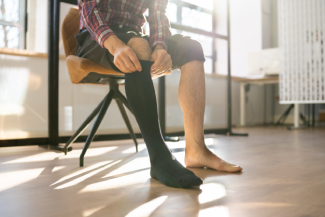
We all know that compression therapy is the “gold standard” for the treatment of venous leg ulcers. However, how do we know if we should apply compression, how much compression, and what type of compression? Lower extremity ulcers are common and affect about 1-2% of all U.S. adults.1 These ulcers are usually classified according to cause, pathogenesis, and treatment type. Approximately 70% of leg ulcers are caused by venous disease, and 20% are caused by arterial insufficiency or mixed arteriovenous disease.2Venous ulcers occur in the gaiter area of the ankle secondary to damaged or dilated veins and retrograde blood flow resulting in venous hypertension. Arterial ulcers result from impaired tissue perfusion.
Are All Compression Bandages the Same?
Compression therapy comes in different types that deliver different amounts of compression. Common types of compression bandages and devices include the following:
- Short stretch or inelastic
- Long stretch or elastic
- Single layer
- Multiple layers
- High and low pressure
Short stretch bandaging systems are more effective at a lower resting pressure than multi-stretch systems. A lower resting pressure offers safer compression in a compromised limb. Both short stretch and multi-stretch systems can produce effective, dynamic working and resting pressures.3
Can compression therapy be used in patients with edema and an ABI of less than 0.8?
All limbs should be examined for adequate arterial supply before applying compression. Patients who do not have palpable pedal pulses should be tested with a Doppler ultrasound to determine their ankle brachial pressure indices (ABIs). If the ABI is above 0.8, the arterial supply is likely to be adequate for full strength 30-40 mmHg compression.3 With an ABI between 0.5 and 0.8, only light compression is to be used. One study showed that patients using a 2 layer light compression system with an ABI of 0.5-0.8 had no pressure-related skin damage, no pain related tissue hypoxia, and reported comfort when wearing the compression device.4 If less than 0.5 ABI, it is recommended that clinicians use compression only with direct supervision by a vascular specialist.5
Can compression therapy be used in a patient with edema and cellulitis?
Cellulitis is an acute bacterial infection of the skin and subcutaneous tissue, most often caused by streptococci or staphylococci.6 Normal antistreptococcal properties of the skin are inactivated by edema fluid. Compression therapy helps to remove protein-containing fluid from the subcutaneous tissues, increases blood flow to those tissues, and thus increases the antibiotic concentration in the tissues. One study by the World Health Organization looked at the efficacy of 8 weeks of antibiotic treatment with compression and without compression. The study found that no patient developed worsening infection or sepsis, and those treated with compression resolved their cellulitis faster than those not treated with compression.7
Does compression therapy improve the skin of patients with venous dermatitis?
Stasis dermatitis results from the insufficient venous return, along with an inflammatory process mediated by metalloproteinases (MMP), which are up-regulated by ferric ions from extravasated red blood cells.8 Edema fluid is more proinflammatory, which allows for higher protease activity.9 Studies have shown that the use of compression therapy decreases the levels of MMPs and cytokines.10 Therefore, the use of compression therapy can help improve venous dermatitis.
Can compression therapy be used in patients with edema and acute deep venous thrombophlebitis?
According to a new study performed by a team of researchers in the Netherlands, individuals with deep-vein thrombosis should start compression therapy within 24 hours of diagnosis. The study showed that patients who received immediate compression therapy saw a reduction of 20% in their risk of developing residual vein occlusion.11 Compression therapy helps to increase venous flow and does not cause an increase in pulmonary embolism. The only contraindication for compression therapy in patients with deep-vein thrombosis would be if their leg is so painful that the patient cannot tolerate the therapy.
References
- Alavi A, Sibbald RG, Phillips TJ, et al. What’s new: management of venous leg ulcers: approach to venous leg ulcers. J Am Acad Dermatol. 2016;74:627-640. https://doi.org/10.1016/j.jaad.2014.10.048 ,>
- Agale SV. Chronic leg ulcers: epidemiology, aetiopathogenesis, and management. Ulcers. 2013; https://www.hindawi.com/journals/ulcers/2013/413604/.
- Raffetto JD, Marston WA. Venous ulcer: What is new? Plast Reconstr Surg. 2011;127(Suppl 1):279S–88S. doi:10.1097/PRS.0b013e3181fcaff2
- Junger M, Hermann H, Ladwig A, et al. Compression therapy in patients with peripheral arterial occlusive disease: a propective clinical study with the 3M coban 2 layer lite compression system for ABPI >0.5. 3M Critical and Chronic Care Solutions Division. 2013; Accessed December 19, 2022. https://multimedia.3m.com/mws/media/909102O/coban-2-abstract-junger.pdf
- Ankle brachial index: quick reference guide for clinicians. Wound, Ostomy and Continence Nurses Society. 2005. Updated 2011. https://www.mision-compresion.es/upload/publicaciones/AnkleBrachialInde…
- Baddour LM. Cellulitis syndromes: an update. Int J Antimicrob Agents. 2000;14: 113–116
- Johnson RC, Sáez-López E, Anagonou ES, et al. Comparison of 8 weeks standard treatment (rifampicin plus clarithromycin) vs. 4 weeks standard plus amoxicillin/clavulanate treatment [RC8 vs. RCA4] to shorten Buruli ulcer disease therapy (the BLMs4BU trial): study protocol for a randomized controlled multi-centre trial in Benin. Trials. 2022;23(1):559. Published 2022 Jul 8. doi:10.1186/s13063-022-06473-9
- Sundaresan S, Migden MR, Silapunt S. Stasis Dermatitis: Pathophysiology, Evaluation, and Management. Am J Clin Dermatol. 2017;18, 383–390. https://doi.org/10.1007/s40257-016-0250-0
- Trengove NJ, Langton SR, MC Stacey MC. Biochemical analysis of wound fluid from nonhealing and healing chronic leg ulcers. Wound Repair Regen. 1996;4(2):234-239. doi:10.1046/j.1524-475X.1996.40211.x
- Marston WA, Beider S, Davies S, et al. Protease and Cytokine Levels in Non-Healing Venous Leg Ulcers Before and After Compression Therapy. Talk Presented at Symposium on Advanced Wound Care/Wound Healing Society Meeting; April 25, 2008; San Diego, CA.
- Amin EE, Bistervels IM, Meijer K, Tick LW, et al. Residual vein occlusion in relation to immediate compression and postthrombotic syndrome in deep vein thrombosis. Blood. 2018; doi: 10.1182/blood-2018-03-836783
About the Authors
Emily Greenstein, APRN, CNP, CWON-AP, FACCWS is a Certified Nurse Practitioner at Sanford Health in Fargo, ND. She received her BSN from Jamestown College and her MSN from Maryville University. She is certified as an Adult-Gerontology Nurse Practitioner through the American Academy of Nurse Practitioners. She has been certified in wound and ostomy care through the WOCNCB for the past 12 years. At Sanford she oversees the outpatient wound care and is co-director for the limb preservation program. She currently serves as the President elect for the North Central Region Wound, Ostomy, and Continence Society. Emily has served as an expert reviewer for the WOCN Society and the Journal for WOCN. Her main career focus is on the advancement of wound care through evidence-based research.
Terry Treadwell, MD, FACS received his medical education at The University of Texas Southwestern Medical School in Dallas, Texas. He continued his training doing his general and vascular surgical residencies at Scott and White Medical Center in Temple, Texas. He practiced vascular and general surgery in Montgomery, Alabama. In October 1998, Dr. Treadwell founded the Institute for Advanced Wound Care at Jackson Hospital in Montgomery. He served as Medical Director of the center and treated wound patients on a full-time basis, providing the best possible care to this seemingly forgotten group of patients. Dr. Treadwell serves as the Medical Director of the Institute.
He has been involved with numerous educational and research initiatives and directs wound care educational programs at his wound center to help educate physicians and other medical personnel on current therapy of acute and chronic wounds. Dr. Treadwell has shared his experience in the treatment of chronic wounds through lectures, presentations, and publications. Wound care practitioners from around the world have attended preceptorships at the Institute for Advanced Wound Care. He has assisted in the establishment of wound treatment centers in Ghana, Africa, and Port-au-Prince, Haiti. He is the Senior Clinical Editor of Wounds magazine and is a member of the World Association of Medical Editors. He is a member of the Wound Healing Society and the Association for the Advancement of Wound Care (AAWC).
The views and opinions expressed in this blog are solely those of the author, and do not represent the views of WoundSource, HMP Global, its affiliates, or subsidiary companies.


Follow WoundSource
Tweets by WoundSource