Corneal Wound Healing
November 11, 2020
Keywords
Categories
By Roshni Patel, BSc (Hons), MCOptom
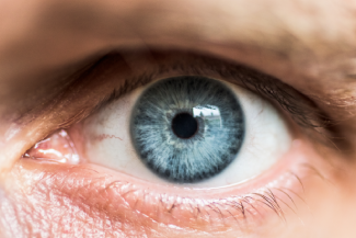
The cornea, as we know it, is a complex and fast-healing tissue that provides protection from infectious and non-infectious defects. However, it can still be injured through environmental impacts or surgical procedures, such as cataract operations or laser-assisted in situ keratomileusis (LASIK), as well as traumatic injuries. Although most corneal wounds repair themselves without further drawbacks,1 possible complications may occur and delay the healing process. This blog outlines the natural healing process of corneal wounds and post-surgical wound healing, as well as the obstacles to healing that may occur in diabetic patients.
Corneal Epithelial Wound Healing
The cornea consists of six layers, from outer to inner: corneal epithelium, Bowman’s layer, stroma, Dua’s layer,2 Descemet’s membrane, and corneal endothelium. Knowing the anatomy of these layers is important to understand the healing process of the cornea:
- Epithelium – The anterior layer of the cornea consists of five to seven layers built on several cell types to maintain the dehydrated condition of the cornea and support the network of materials that are responsible for the cornea’s permeability. Cells of the epithelium are renewed approximately every 10 days to allow corneal injuries to heal quickly.
- Bowman’s layer – This layer is acellular and mainly consists of collagen arranged in fibrils that interweave with the stromal layer below. This layer is highly resistant to damage, but it is unable to regenerate once compromised.
- Stroma – The stromal layer consists of collagen fibrils, proteoglycan aggregates that create a porous hydrated gel, and keratocytes as the primary cell type. The latter produces glycosaminoglycans (GAGs) and matrix metalloproteinases (MMPs). Together, this network is responsible for the integrity and thickness of the cornea.
- Dua’s layer – This is a thin acellular layer consisting of five to eight collagen bundles in diagonally crossed arrangements. It is free of keratocytes but spaced in a similar pattern to the stromal layer, in contrast to the tight network of the Descemet’s membrane. Dua’s layer is especially tough and resilient to pressure.
- Descemet’s membrane – Descemet’s membrane consists of collagen in the anterior layer to provide elasticity and the posterior layer is created by secretion of the endothelium. This layer is, similarly to the Bowman’s layer, highly resistant, yet unable to regenerate once damaged.
- Endothelium – The final endothelial layer maintains the deturgescence of the cornea because it consists of one singular layer of polyhedral cells. Its cells are organized in a network of tight junctions and gap junctions that create a penetrable surface to allow the cornea to intake nutrients from the aqueous humor. The cells of the endothelium are unable to replicate or divide; therefore, they will change in size to fill spaces of lost cells.
Latent Phase
This phase of the epithelial healing process takes place over the first few hours following the injury and is characterized by the reshaping of cells. The damaged cells undergo apoptosis and are shed into the tear film, and they are subsequently removed from the ocular surface. The network built from adherens junctions and gap junctions surrounding the injury is lost, and the attached basal cells are broken down by the rim of the wound. They continuously change in shape and form the cellular projections filopodia and lamellipodia to serve as a foundation for epithelial cells in close proximity to move over the wound.
Migration Phase
During this phase, cells move across the affected area in a single layer. The cell extensions filopodia and lamellipodia form temporary attachments to the underlying material and glide across the wound’s edge, where the attachments are dropped. This cycle continues for approximately 24 to 36 hours, depending on the size and location of the wound, until it is completely covered.
Proliferation Phase Following the migration, the single layer of cells is extended to reconstruct the regular thickness of the epithelium layer. This is achieved by means of basal cells that convert over wing cells to form squamous cells during movement. To repair the barrier function of the cornea, tight junctions are formed, and desmosomes, gap junctions, and adherens junctions are restored between the cells.
Epithelial Reattachment
This final phase is the re-anchoring of cells, in which hemidesmosomes function as anchoring fibrils to attach the epithelial layer to the substrate. If the basement membrane did not incur any damage, this process usually occurs over a period of days. However, if the damage reaches the basement membrane, the healing process will stretch over several months.
Post-Surgical Corneal Wounds
Thanks to its complex structure, the cornea is resistant to the majority of environmental impacts. However, some routine ocular surgeries cause corneal wounds through small incisions.
Cataract Surgery
Phacoemulsification is a routine procedure for ophthalmologists. After centuries of development, the preferred method of accessing the cataract nowadays is often through a clear corneal incision4 because it reportedly has a lower chance of causing conjunctival trauma, is more comfortable for the patient, and causes less bleeding, thus resulting in a swift recovery of the patient’s visual acuity. It uses clear corneal wounds, allowing not only for a shorter surgical process but also reducing the recovery period after the surgery. However, there are a few cases of corneal wound infections after phacoemulsification using a clear corneal incision, such as wound burning,5 caused by heat generated by the phacoemulsification tip during surgery. This infection can be recognized by a decreased flow of lens milk and stagnant emulsification, and it can be treated by means of bandage contact lenses and aqueous suppressants.
Laser-Assisted In Situ Keratomileusis
Although glasses and contact lenses6 are reliable temporary options for the correction of refractive errors, some patients may choose LASIK to correct their myopia or astigmatism permanently. In this surgical procedure, which is considered safe and effective, the corneal flap7 is cut with an oscillating blade, and a laser reshapes the stromal layer by removing tissue before the flap is placed back onto the ocular surface. It does not rely on the large myofibroblast8 cells, which are reported to cause post-surgical corneal scarring and corneal haze. Although LASIK is a routine procedure and usually causes only minimal inflammation, deficiencies such as an epithelial defect, lamellar keratitis, or flap folds may hinder the healing process on rare occasions. Over the last decade, corneal ectasia has increasingly been reported as a consequence of LASIK, in some cases caused by a form of abnormal wound healing.9 This defect can occur one week after LASIK but it may develop several years after surgery. It is often misdiagnosed as post-LASIK keratoconus.
Keratoplasty
Scarred or deceased corneal tissue, either through injury or disease, can be replaced through a corneal transplant10 from an organ donor, either by penetrating keratoplasty (PK) or endothelial keratoplasty (EK). During PK transplants, a trephine or femtosecond laser is used to remove the affected corneal areas, before a matching area of the donor tissue is positioned and structured into place through sutures. During the healing process, a plastic shield is placed over the eye to prevent any infection. The more recently introduced process of EK is used for specific corneal conditions. The endothelial particles of the donor tissue are applied through a small, self-sealing incision. The possible complications of keratoplasty can be caused by rejection of the donor tissue or, more commonly, surgical wound dehiscence.11
Diabetic Corneal Wounds
The process of corneal wound healing is complex, and although most of these wounds heal without complications, patients with diabetes are more susceptible to experiencing impaired corneal wound healing. Several studies show that various factors can cause complications in corneal wound healing in diabetic patients, including the weaker production of wound electric signals and higher glucose levels, as well as increased oxidative stress in the corneal epithelium. This can lead to corneal ulcers, causing further difficulties.
The treatment of diabetic corneal wounds continues to evolve. One way that physicians can treat these corneal epithelial wounds is to administer a synergy peptide Pro-His-Ser-Arg-Asn (PHSRN)12 topically in the form of eye drops to expedite the healing process. Further studies are needed to clarify the different causes of corneal diabetic defects and to find therapeutic methods that offer better and results.
References
1. Cornea. Healthline. https://www.healthline.com/human-body-maps/cornea#1. Accessed September 21, 2020.
2. Sixth layer to human cornea discovered. Optometry Times. 2013. https://www.optometrytimes.com/view/sixth-layer-human-cornea-discovered. Accessed September 21, 2020.
3. Lee TN. The ins and outs of corneal wound healing. Review of Optometry. 2016. https://www.reviewofoptometry.com/article/the-ins-and-outs-of-corneal-w…. Accessed September 21, 2020.
4. Al Mahmood AM, Al-Swailem SA, Behrens A. Clear corneal incision in cataract surgery. Middle East Afr J Ophthalmol. 2014;21(1):25-31. https://www.ncbi.nlm.nih.gov/pmc/articles/PMC3959037/. Accessed September 21, 2020.
5. Jacob S. Complications of cataract surgery. EuroTimes. 2018. https://www.eurotimes.org/eye-wound-burn-complications-jacob/. Accessed September 21, 2020.
6. Contact lenses. Lenstore.co.uk. https://www.lenstore.co.uk/contact-lenses. Accessed September 21, 2020.
7. What is the ‘flap’ in laser eye surgery? Vision Eye Institute. 2017. https://visioneyeinstitute.com.au/eyematters/flap-laser-eye-surgery/. Accessed September 21, 2020.
8. Riau AK, Angunawela RI, Chaurasia SS, Lee WS, Tan DT, Mehta JS. Early corneal wound healing and inflammatory responses after refractive lenticule extraction (ReLEx). Invest Ophthalmol Vis Sci. 2011;52(9):6213-6221. https://iovs.arvojournals.org/article.aspx?articleid=2187266. Accessed September 21, 2020.
9. Dupps WJ Jr, Wilson SE. Biomechanics and wound healing in the cornea. Exp Eye Res. 2006;83:709-720. https://www.ncbi.nlm.nih.gov/pmc/articles/PMC2691611/. Accessed September 21, 2020.
10. Boxer Wachler BS. Cornea transplants: what to expect from keratoplasty. All About Vision. https://www.allaboutvision.com/conditions/cornea-transplant.htm. Accessed September 21, 2020.
11. Jafarinasab MR, Feizi S, Esfandiari H, Kheiri B, Feizi M. Traumatic wound dehiscence following corneal transplantation. J Ophthalmic Vis Res. 2012;7(3):214-218. https://www.ncbi.nlm.nih.gov/pmc/articles/PMC3520589/. Accessed September 21, 2020.
12. Bu Y, Shih KC, Kwok SS, et al. Experimental modeling of cornea wound healing in diabetes: clinical applications and beyond. BMJ Open Diabetes Res Care. 2019;7(1):e000779. https://pubmed.ncbi.nlm.nih.gov/31803484/. Accessed September 21, 2020.
About the Author
Roshni Patel, BSc (Hons), MCOptom, qualified as an Optometrist in 2004. She is a member of the Association of Optometrists and is the Professional Services Manager at Lenstore, where she is responsible for supporting and guiding all departments from a clinical standpoint.
The views and opinions expressed in this content are solely those of the contributor, and do not represent the views of WoundSource, HMP Global, its affiliates, or subsidiary companies.