Identifying Types of Tissues Found in Pressure Ulcers
November 20, 2014
Keywords
Categories
By the WoundSource Editors
Over the course of a wound’s existence, several tissue types can be identified. In order to properly stage a pressure ulcer (injury) and determine the best treatment option, it is important for the clinician to be able to determine the tissue type that is present. The following represent the most commonly identified tissue types seen in pressure ulcers, and also in other open wounds:
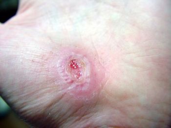
Epithelium
Epithelial tissue often appears lighter than surrounding tissue (i.e. light pink in color). Epithelialization occurs when the epidermis regenerates over a wound surface. Basal keratinocytes travel from the wound edges, where they multiply until they meet in the middle. The basal lamina is a scaffolding secreted by the epithelial cells as they travel outwards from the wound edges.
When a new epithelial layer is created, this new layer is only a few cell layers in thickness and appears translucent. It is extremely vulnerable to damage from friction, shearing and pressure. It generally takes 2 to 3 weeks for the new cells to become keratinized (i.e. waterproof).
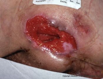
Granulation Tissue
The formation of granulation tissue is thought to be an intermediate step in the healing process of full-thickness wounds. Granulation tissue is also very fragile and prone to easy injury. Granulation tissue is shiny red and granular in appearance when it is healthy; when inadequate blood flow exists, granulation tissue may pale in color. The process of granulation provides the early scaffolding necessary to promote healing from the edges of the wound. Granulation tissue does not mature into epithelium; instead, granulation tissue is eventually covered by a layer of epidermal tissue.
Granulation tissue is subject to injury by outside forces, including dry/adherent dressings, pressure, high-intensity irrigation of the wound and overzealous wound packing. Impaired blood flow and excessive pressure can also damage granulation tissue and may cause patients to complain of new-onset pain when dressings are changed. Wound healing may stall in the granulation phase when nutrients are inadequate, infection is present or blood flow is impaired.
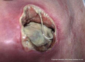
Slough
Slough is indicative of full-thickness stage III pressure ulcers (stage 3 pressure injury) or stage IV pressure ulcers (stage 4 pressure injury). Slough can easily be confused with normal anatomical tissues such as tendons or ligaments because of their frequently yellowish coloration. This can be a costly mistake, due to the fact that slough is non-viable tissue and requires debridement. When a large amount of slough is present and obscures the wound bed, the wound is unstageable.
Slough can be identified as a stringy mass that may or may not be firmly attached to surrounding tissue. Slough can range in color from white (scant bacterial colonization) to yellow or green (larger bacterial counts) to brown (hemoglobin is present). Slough may become thicker and harder to remove the longer it is present.
Eschar
Eschar is composed of necrotic granulation tissue, muscle, fat, tendon or skin. The term stable eschar is used to describe leathery, dry hard eschar tissue, such as the eschar that commonly forms on the heels or other bony prominences of the lower leg of patients with ischemic limbs.
The term unstable eschar is used to describe tissue that is undergoing a softening process caused by proteolytic enzyme production from bacteria present in the tissues. This type of eschar is characterized by pain, redness, purulent discharge, warmth and edema. This type of eschar tissue may be described as spongy, boggy or slimy. The presence of unstable eschar raises the risk of sepsis, amputation and systemic infection. Wet gangrene should be ruled out when fluctuance, crepitance or purulent drainage is present. Sometimes, autolytic debridement or topical enzymes are used on eschar tissue, and this deliberate softening should not be confused with unstable eschar.
Pressure Ulcer Tissue Assessment for Treatment and Healing Status
As can be seen, each of these tissue types is distinct, with identifiable characteristics and treatment considerations. Professionals involved in describing and treating pressure ulcers must be able to differentiate among epithelium, granulation tissue, slough and eschar in order to ensure that pressure wounds are treated accordingly and safely. Tissue identification is also important to note in the healing progress of a wound. The National Pressure Ulcer Advisory Panel’s (NPUAP) Pressure Ulcer Scale for Healing (PUSH) Tool offers clinicians a way to assess a wound’s status change by scoring pressure ulcers on a number of characteristics, including tissue types present.
References
Ayello E, Baranoski S, Lyder C, Cuddigan J. Chapter 13: Pressure Ulcers. In: Baranoski S, Ayello E, eds. Wound Care Essentials: Practice Principles. Ambler, PA: Lippincott: Williams and Wilkins;2004:240-270.
Black J, Baharestani M, Black S, et al. (2010). An overview of tissue types in pressure ulcers: A consensus panel recommendation. Ostomy Wound Management. 2010;56(4):28-44.
NPUAP. PUSH Tool 3.0 (web version). NPUAP.org. http://www.npuap.org/resources/educational-and-clinical-resources/push-…. Published September 15, 1998. Accessed October 12, 2014.
Editor's Note: This article was originally published November, 2010 and has been updated for accuracy and comprehension.
The views and opinions expressed in this blog are solely those of the author, and do not represent the views of WoundSource, HMP Global, its affiliates, or subsidiary companies.

Follow WoundSource
Tweets by WoundSource