
Pressure Injury and Microclimate: How Linen Layers May Contribute
Laura Swoboda, DNP, APNP, FNP-C, FNP-BC, CWOCN-AP, WOCNF
February 14, 2024
February 14, 2024
Keywords
Categories
Introduction
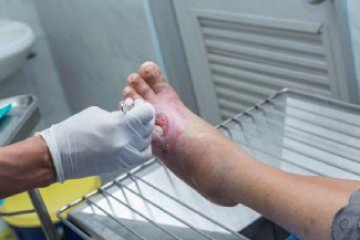
Pressure injuries (PI) and their effect on patients across settings are a growing concern for wound care professionals. Of note, patients with pressure injuries admitted to the ICU have a significantly higher risk of mortality as compared to patients without PIs.1 Strategies to prevent and manage these injuries are constantly explored by leaders in the field, with special attention to linens and dressings.1,2 Linens are frequently placed under hospitalized patients for a multitude of reasons including3:
- Moisture management
- Incontinence containment
- Repositioning of immobile patients
Hospital mattresses and specialty support surfaces can offload pressure and manage moisture, but the number and type of additional layers between the patient and support surface may affect efficacy. For example, incontinence containment pads can consist of quilted fabric, paper backing, or plastic elements, all with varying outcomes. Subtypes of layers between the patient and the support surface may impact the skin microclimate.3
What Is the Skin Microclimate?
For this discussion, the dermal microclimate is where the patient’s skin contacts their support surface. Just as an environmental microclimate is a local set of atmospheric conditions that differ from surrounding areas, the skin microclimate differs from other areas of the body.1,2 It is known that pressure injury risk is impacted by a constellation of features, including those that regulate microclimate. Factors related to skin microclimate that affect tissue tolerance include4,5:
- Moisture (Humidity)
- Temperature
- Air movement
By significantly impacting the skin and its supporting structures, it is proposed that the skin is less able to endure pressure, friction, and shear without adverse sequelae.6 In particular, when skin microclimate rises, the cutaneous layers of the skin lose transepidermal water, increasing moisture on the skin’s surface and reducing the strength of the stratum corneum.6 The risks created by the skin microclimate constitute a collection of modifiable indirect pressure injury risk factors. Although these factors are modifiable, an exact recommendation for skin microclimate remains undetermined among experts.6
Pressure Injury Risk: How Linen Layers May Contribute
Linen layers have the potential to impact skin microclimate in the following ways7:
- Reduce airflow
- Affect pressure redistribution
- Increase friction coefficient
- Dry or macerate skin
- Increase skin temperature
Clinicians should select the correct type and amount of layers between the patient and support surface. Evidence suggests this selection is a high-value, low-cost, intervention.1-15 Regarding linen type, the NPIAP 2019 guidelines specifically recommend the use of silk or silk-like sheets versus cotton and cotton blend sheets.2 Silk or silk-like linens have been found to have a lower friction coefficient and quicker drying times as compared to cotton-based linens.2
Regarding linen number, experts have found that incontinence pads, transfer sheets, or a combination of linens can significantly increase the mean peak sacral pressure when compared to a single flat sheet.8 Even on both a low-air-loss surface and foam surface, regardless of head-of-bed angle, pressure may be increased.8 This occurrence was confirmed by an in vitro study by Fader et al which examined the effect on interface pressures with the use of wet and dry incontinence pads against the gluteal and sacral areas of mannequins with soft, tissue-like qualities.9 In a 2018 retrospective review using International Pressure Ulcer Prevalence data from 216,626 participants, additional linen layers were found as a risk factor for both superficial and severe pressure injuries.10
Multiple pressure injury prevention programs recommend reducing linen layers, including guidelines regarding ICU patients.11 The NPIAP 2019 guidelines acknowledge the need for incontinence pads and positioning devices that are compatible with support surfaces.2 They provide specific directions to limit the amount of linen and pads placed on the bed.2
Reviewing the Evidence Further
As previously stated, how linens and pads contribute to pressure injury development usually depends on how they affect microclimate factors, including moisture amount. A 2019 study exposed 12 subjects on pressure-loaded surfaces to moist incontinence pads.12 The researchers found increased transepidermal water loss and upregulated inflammatory markers in the subjects exposed to moist pads, as opposed to subjects exposed to dry pads who experienced no transepidermal water loss.12
Incontinence-associated dermatitis (IAD) is another indirect risk factor for pressure injury development. A 2016 retrospective analysis of pressure injury prevalence survey data found additional linen layers were associated with the development of IAD, increasing overall risk for IAD by 8.3%.13-15 A similar study investigated the use of one layer of plastic-containing underpads. It found that the underpad’s use significantly reduced the surface’s ability to modulate microclimate, with reductions in heat dissipation and moisture evaporation.8
Conclusion
A review of literature clearly confirms that excess and certain linen layers can increase the risk of pressure injury by directly increasing interface pressure between the patient and the support surface as well as through reducing a therapeutic surface's ability to manage the dermal microclimate.2-15
References
- Montague-McCown M, Bena J, Burchill CN. Effect of Hospital Linens on Unit-Acquired Pressure Injuries for Adults in Medical ICUs: A Cluster Randomized Controlled Trial. Crit Care Explor. 2021;3(3):e0336.
- European Pressure Ulcer Advisory Panel, National Pressure Injury Advisory Panel and Pan Pacific Pressure Injury Alliance. Prevention and Treatment of Pressure Ulcers/Injuries: Clinical Practice Guideline. Emily Haesler (Ed.). EPUAP/NPIAP/PPPIA: 2019
- Williamson R, Lachenbruch C, Vangilder C. The effect of multiple layers of linens on surface interface pressure: results of a laboratory study. Ostomy Wound Manage. 2013;59(6):38-48.
- Bauer K, Rock K, Nazzal M, et al. Pressure ulcers in the United States' inpatient population from 2008 to 2012: results of a retrospective nationwide study. Ostomy Wound Manage. 2016;62(11):30-8.
- Bryant R, Nix D. Acute & Chronic Wounds: Current management concepts, fourth edition. Elsevier; 2012. IBSN: 9780323069434
- Borzdynski C, Miller C, Vicendese D, McGuiness W. Brief intermittent pressure off-loading on skin microclimate in healthy adults - a descriptive-correlational pilot study. J Tissue Viability. 2021; 30(3):379-394. https://www.sciencedirect.com/science/article/pii/S0965206X21000322
- Edsberg LE, Cox J, Koloms K, VanGilder-Freese CA. Implementation of Pressure Injury Prevention Strategies in Acute Care: Results From the 2018-2019 International Pressure Injury Prevalence Survey. J Wound Ostomy Continence Nurs. 2022;49(3):211-219. doi:10.1097/WON.0000000000000878
- Williamson R, Lachenbruch C, VanGilder C. A laboratory study examining the impact of linen use on low-air-loss support surface heat and water vapor transmission rates. Ostomy Wound Manage. 2013;59(8):32–41.
- Fader M, Bain D, Cottenden A. Effects of absorbent incontinence pads on pressure management mattresses. J Adv Nurs. 2004;48(6):569-574. doi:10.1111/j.1365-2648.2004.03245.x
- Kayser SA, VanGilder CA, Lachenbruch C. Predictors of superficial and severe hospital-acquired pressure injuries: a cross-sectional study using the International Pressure Ulcer Prevalence survey. Int J Nurs Stud. 2018;89:46–52.
- Krupp AE, Monfre J. Pressure ulcers in the ICU patient: an update on prevention and treatment. Curr Infect Dis Rep. 2015;17(3):468.
- Bostan LE, Worsley PR, Abbas S, Bader DL. The influence of incontinence pads moisture at the loaded skin interface. J Tissue Viability. 2019;28(3):125-132. doi:10.1016/j.jtv.2019.05.002
- Kayser SA, Phipps L, VanGilder CA, Lachenbruch C. Examining Prevalence and Risk Factors of Incontinence-Associated Dermatitis Using the International Pressure Ulcer Prevalence Survey. J Wound Ostomy Continence Nurs. 2019;46(4):285-290. doi:10.1097/WON.0000000000000548
- Demarre L, Verhaeghe S, Van Hecke A, Clays E, Grypdonck M, Beeckman D. Factors predicting the development of pressure ulcers in an at-risk population who receive standardized preventive care: secondary analyses of a multicentre randomised controlled trial. J Adv Nurs. 2015;71(2):391–403.
- Gray M, Giuliano KK. Incontinence-associated dermatitis, characteristics and relationship to pressure injury: a multisite epidemiologic analysis. J Wound Ostomy Continence Nurs. 2018;45(1):63–67.
About the Author
Dr. Laura Swoboda, DNP, APNP, FNP-BC, CWOCN-AP, is a Professor of Health Sciences, Family Nurse Practitioner, and certified wound specialist in Milwaukee Metro, Wisconsin. Dr. Swoboda is on the NPIAP’s Prophylactic Dressing Standards Initiative Task Force, a member of the editorial board for the Wound Care Learning Network and Wound Management and Prevention, and on the board of directors for the WOCNCB and the AAWC.
The views and opinions expressed in this blog are solely those of the author, and do not represent the views of WoundSource, HMP Global, its affiliates, or subsidiary companies.


Follow WoundSource
Tweets by WoundSource