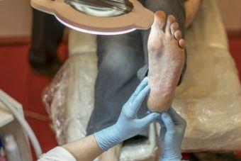
Complex Wound Management: Diabetic Foot Ulcers
August 27, 2018
Background and Prevalence of Diabetic Foot Ulcers
Diabetes-related foot complications, including diabetic foot ulcers (DFUs), are leading causes of non-traumatic lower extremity amputation. Of the approximately 420 million adults in the United States with diabetes mellitus, one fourth will develop at least one DFU.1,2 DFUs are preceded by a compendium of risk factors, including the presence of neuropathy, external trauma, infection, effects of ischemia from concomitant peripheral arterial disease, malnutrition, and poor hygiene or self-care, among others. In 80% of patients, DFU is a precursor to some degree of lower extremity amputation.3 And, for these patients who have undergone amputation, their risk for further amputation becomes double that of a patient without diabetes. The mortality rate following a diagnosis of diabetic foot ulceration is 5% in the first year. The five-year mortality rate is 50% and rises to 70% after amputation.4 Once healed, 40% of DFUs will recur within 12 months, nearly 70% at three years, and nearly 75% at five years.5
Evidence-Based Management of Diabetic Foot Ulcers
- Disease Management: Medical management and chronic disease optimization for individuals with diabetic foot ulceration includes glucose control by dietary control and/or insulin or oral hypoglycemic agents, management of hypertension if present, fluid volume control (relative to lower extremity edema), and adequate nutrition, along with any supplements appropriate for wound healing.
- Wound Assessment: Routine wound assessments should be recorded using standardized routine documentation to best track healing progress and include measurements and wound characteristics with any noted signs of infection, grading and classification of DFU for risk stratification and treatment purposes, and presence of ischemia, neuropathy or arthropathy. Repeat assessments should include reclassification of the DFU, if necessary.
- Wound Bed Preparation: Serial sharp excisional debridement of nonviable tissue will ideally include excision of periwound callus and callused wound edge.
- Topical Wound Management: Topical therapy is inclusive of appropriate dressings tailored to dynamic wound characteristics, with attention to moisture and exudate levels. Advanced therapies include negative pressure wound therapy (NPWT), cellular and/or tissue-based products (CTPs), growth factors (GFs), and hyperbaric oxygen therapy (HBOT) as indicated. NPWT for DFU decreases length of hospital stay and complication rates. Numerous CTPs are available; some evidence suggests a decreased risk for amputation in reviewed trials and an improved rate of closure compared with standard care.7 Evidence suggests higher rates of DFU closure with use of GFs, notably platelet-derived GF (PDGF).8 HBOT studies show decreased time to closure and reduced chance of amputation in DFUs in which at least 30 days of standard therapy have failed in patients with improved transcutaneous oxygen pressure (TcPO2 tissue oxygenation) testing after HBOT.9 There is a trending use of these adjunctive advanced therapies with the increase in research, clinicians’ training and comfort with therapies, clinicians’ education, and specialization in wound care. No ubiquitous consensus has been reached on recommendation of these therapies for regular use in routine care of the DFU.
- Offloading Interventions: Mechanical offloading is a cornerstone of DFU management because the pathology and etiology of the disease process are perpetuated by pressure and friction from altered biomechanics of the foot. Total contact casting, controlled ankle movement (CAM) walkers, footwear modifications, and other devices may be used for offloading. Contact casting is the gold standard for offloading of DFU when a clinician skilled in the application of this technique is available.10 In the absence of an experienced clinician, removable CAM walkers may be preferable.
- Edema Management: Edema management includes the application of lower extremity compression if appropriate, coupled with elevation of the extremity above the level of the heart when the patent is not ambulating.
- Surgical Interventions: Surgical correction may be required for bony deformities that may by difficult to offload or those refractory to attempts at mechanical offloading (e.g., prominent metatarsal head with overlying ulceration, hammer toes with ulceration at the distal tip of the toe).
- Infection Management: Treatment of infection includes confirmation of depth of infection and superficial soft tissue versus osteomyelitis or sepsis (based on bioburden continuum). A tissue specimen should be obtained after cleansing and debridement and sent for qualitative culture and sensitivities. If empiric antibiotics are required to treat spreading infection, it is ideal to obtain the culture before antibiotics are initiated. Swab or exudate cultures are not considered standard of care.
- Vascular Interventions: Revascularization of ischemic limbs is associated with a lower incidence of amputation in the presence of a DFU.
- Education: Patient education of disease processes and rationale for interventions related to treatment of DFU is integral because outcomes are directly related to the patient’s knowledge of his or her disease process and self-care ability.3
These recommendations are aligned with the Infectious Disease Society of America 2012 practice guidelines and the 2016 joint guidelines of the Society for Vascular Medicine and the American Podiatric Medical Association. Multidisciplinary care is crucial for patients with DFU, and it should be carried out by a multidisciplinary team, with the previously reviewed items included as a total plan of care. Even after a patient’s ulceration is resolved, the patient is at high risk for DFU recurrence. Some literature has begun to refer to this resolution as “remission,” which better captures the concept that ulceration may recur. Long-term goals are to engage the patient in adequate preventive care and encourage the patient’s maximum activity levels.11
References
1. Alavi A, Sibbald RG, Mayer D, et al. Diabetic foot ulcers: part II. Management. J Am Acad Dermatol. 2014;70(1):21.e1–24; quiz 45.
2. World Health Organization. Global Report on Diabetes. Geneva, Switzerland: World Health Organization; 2016. http://apps.who.int/iris/bitstream/handle/10665/204871/9789241565257_en…. Accessed August 6, 2018.
3. Chadwick P, Edmonds M, McCardle J, Armstrong D. International best practice guidelines (IBPG): wound management in diabetic foot ulcers. Wounds International. 2013. http://www.woundsinternational.com/best-practices/view/best-practice-gu…. Accessed August 6, 2018.
4. National Institute for Health and Care Excellence. Type 2 diabetes in adults: management. NICE guideline NG 28. London, United Kingdom: National Institute for Health and Care Excellence; 2015. https://www.nice.org.uk/guidance/ng28. Accessed August 6, 2018.
5. Armstrong DG, Boulton AJM. Diabetic foot ulcers and their recurrence. N Engl J Med. 2017;376(24):2367–75.
6. Driver VR, Fabbi M, Lavery LA, et al. The costs of diabetic foot: the economic case for the limb salvage team. J Vasc Surg. 2010;52(3 Suppl):17S–22S.
7. Santema TB, Poyck PP, Ubbink DT. Skin grafting and tissue replacement for treating foot ulcers in people with diabetes. Cochrane Database Syst Rev. 2016;(2):CD011255.
8. Martí-Carvajal AJ, Gluud C, Nicola S, et al. Growth factors for treating diabetic foot ulcers. Cochrane Database Syst Rev. 2015;(10):CD008548.
9. Alavi A, Sibbald RG, Mayer D, et al. Diabetic foot ulcers: part I. Pathophysiology and prevention. J Am Acad Dermatol. 2014;70(1):e1–18.
10. Spencer S. Pressure relieving interventions for preventing and treating diabetic foot ulcers. Cochrane Database Syst Rev. 2000;(3):CD002302.
11. Hingorani A, LaMuraglia GM, Henke P, et al. The management of diabetic foot: a clinical practice guideline by the Society for Vascular Surgery in collaboration with the American Podiatric Medical Association and the Society for Vascular Medicine. J Vasc Surg. 2016;63(2 Suppl):3S–21S.
The views and opinions expressed in this content are solely those of the contributor, and do not represent the views of WoundSource, HMP Global, its affiliates, or subsidiary companies.


