Skin Grafting of Surgical Wounds
February 19, 2013
Keywords
Categories
By Laurie Swezey RN, BSN, CWOCN, CWS, FACCWS
Skin grafting of surgical wounds is performed for wounds that are difficult to close using traditional closure methods, such as staples or sutures. They may also be used for wounds that are expected to result in severe scarring, which may have psychological or physical repercussions for the patient. Skin grafting serves three main purposes: it covers the wound, minimizes scarring and speeds healing.
What is a Skin Graft?
A skin graft is a portion of epidermis and dermis that has been removed from one site (the donor site) on a patient’s body and is transplanted to another site (the recipient site). Skin grafting results in two wounds rather than one, with both having the potential to become infected. Autografts are the most common grafts used, where the donor and recipient is the same patient.
Much like burns, autografts may be full-thickness or partial-thickness (split-skin), depending on how much of the dermis the surgeon chooses to use. A partial-thickness skin graft will involve removal of the epidermis and a portion of the dermis, leaving behind the deep dermis so that the skin can regenerate itself. Full-thickness skin grafts use the full thickness of the dermis. The skin cannot regenerate itself because the reticular dermis is removed. Therefore, the wound is closed by the surgeon to allow for healing by primary intention.
The upper arm, buttock, abdominal wall, back and thigh are common donor site areas in partial-thickness skin grafts. Theskin in front of and behind the ear, antecubital area and supraclavicular area, as well as the groin, scalp, eyelid and areola are areas most commonly used in full-thickness skin grafts.
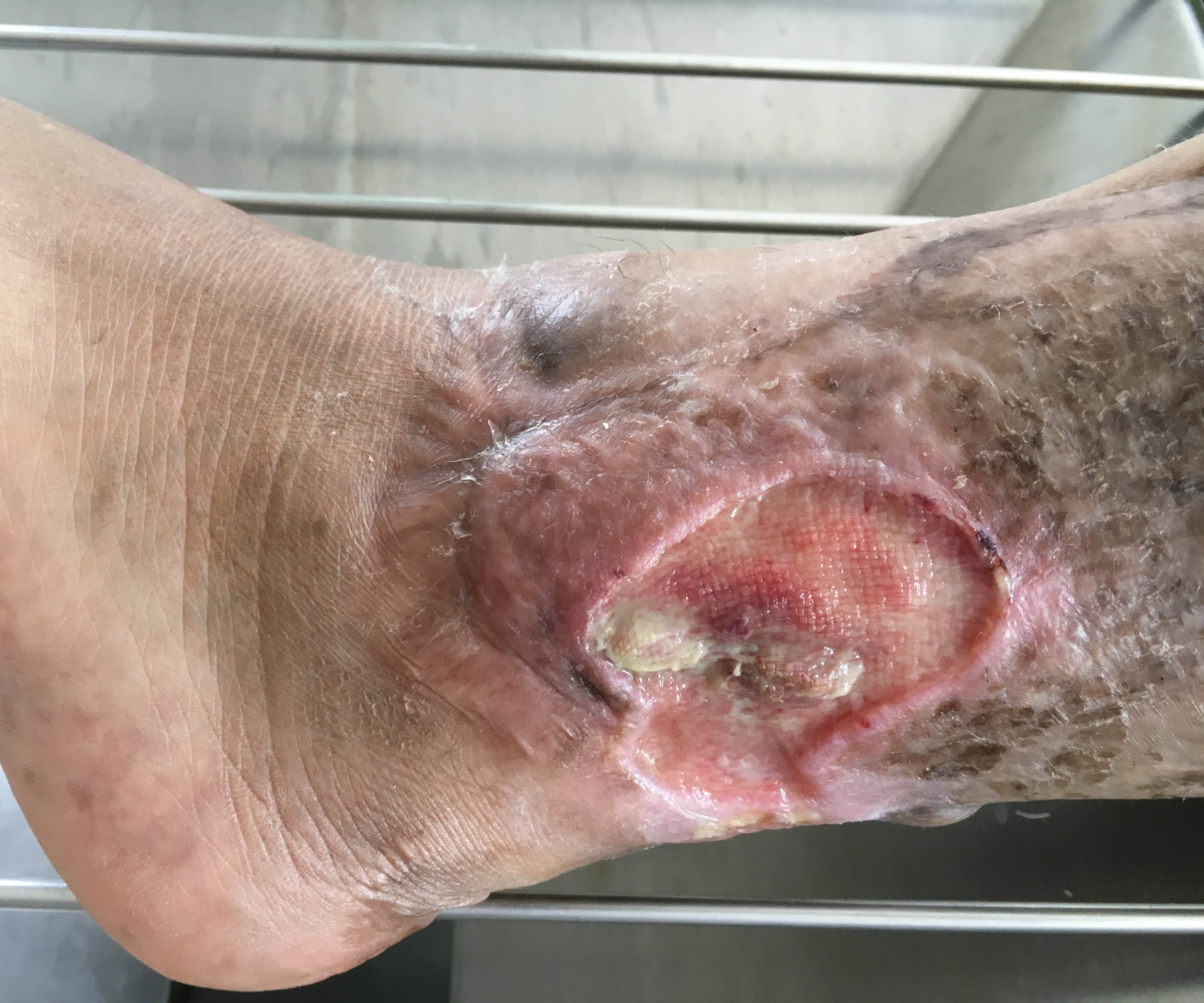
Skin grafts are typically left alone for two to five days following the grafting procedure. Dressings should be removed with care. The graft is then examined to determine whether it has successfully adhered to the wound bed. If healthy, it should be pink or red in color and should be firmly attached to the underlying wound bed. Staples and sutures are removed at this time so that they don’t delay healing. The type of dressing applied depends on how well the graft has adhered to the wound and whether infection is present. An absorbent dressing may be required if the skin grafts site produces large amounts of exudate.
The donor site wound also requires attention and is often more painful than the graft site because sensory nerve endings are exposed. Healing usually occurs within seven to ten days but may take as long as three weeks. In the first few days the donor wound site will produce exudate in varying amounts.
Once the donor site has healed, the skin may be dry and itchy. Patients should be encouraged not to scratch to prevent breaking the skin. Applying emollients may be helpful. Patients should be counseled to apply sunscreen to the area when exposed to the sun for at least the first year after grafting.
Source:
Belden, P. What you need to know about skin grafts and donor site wounds: Technical guide. Wound Essentials, vol 2. 2007 Available at: http://www.woundsinternational.com/practice-development/what-you-need-t…
About The Author
Laurie Swezey RN, BSN, CWOCN, CWS, FACCWS is a Certified Wound Therapist and enterostomal therapist, founder and president of WoundEducators.com, and advocate of incorporating digital and computer technology into the field of wound care.
The views and opinions expressed in this blog are solely those of the author, and do not represent the views of WoundSource, Kestrel Health Information, Inc., its affiliates, or subsidiary companies.
The views and opinions expressed in this content are solely those of the contributor, and do not represent the views of WoundSource, HMP Global, its affiliates, or subsidiary companies.