Weird Wounds Part 2: Calciphylaxis – "The Heart Attack of the Skin"
April 9, 2020
Keywords
Categories
Hypothetical Case Study
Picture this: you've been seeing a patient in your wound center for the last several months to treat a slowly healing post-operative abdominal wound. The wound has been gradually responding to an assortment of treatments, including initial wound vacuum therapy after the surgery, followed by alginate and now a collagen dressing. The wound is getting smaller and has new granulation tissue at the base.
You're actually a bit surprised that it's healing so nicely because the patient has multiple serious chronic illnesses, including severe chronic kidney disease that requires hemodialysis sessions three times per week, type 2 diabetes, morbid obesity, cardiovascular disease, and peripheral vascular disease. One day, the patient missed her appointment, which was fairly unusual for her, so you log into her inpatient record and find that she had been admitted for sepsis two days earlier after developing a high fever. You read that she is being treated with intravenous fluids and intravenous antibiotics, and she seems to be responding well. It seems the plan is for her to be discharged to a nearby rehabilitation facility in the next several days. You hope she recovers without incident, and you continue with your day.

Several weeks after this, she returns to the clinic. You are dismayed to find several new wounds: one on her left heel and the posterior left calf, two wounds on each iliac crest, and one wound on her right medial thigh. The original abdominal wound hasn't changed significantly. She tells you that the new wounds developed while she was in the hospital and they are very painful, especially with dressing changes and with any kind of direct pressure. She describes the pain as "a terrible burn."
You observe that she has alginate dressings and gauze on the wounds. She also tells you that she was on a pressure-relieving air mattress while in the hospital and the rehabilitation facility. She had access to a supportive recliner in her room, but because she was quite weak, she found it difficult to get out of bed without help so spent a large part of her time in bed. She is now home with her family and feeling better. She states that she feels a bit stronger and is able to walk around her home with a walker. She has VNA support for her wound and medication management and has physical and occupational therapists coming to her home twice a week. She continues with her hemodialysis sessions three times per week. She drinks diabetic protein supplement shakes three times a day with each meal.
Despite this, she has lost about 60 pounds over the last three months. When asked about this, she states, "I just don't seem to have my appetite anymore." At first glance, the new ulcers appear to be related to pressure. Certainly, the ulcers on her heel and hips would be at risk because of her prolonged bedrest. However, the calf and medial thigh are strange locations for pressure ulcer formation. Perhaps, in an attempt to offload pressure from the left heel, a pillow or some other kind of device was propped under her calf, creating pressure to this site. And you suppose that it's possible that some kind of wedge or other device was propped between her legs and caused the medial thigh ulcer to develop. You know the patient is very frail, so you think although this would be unusual, it could be possible. You ask the patient these questions, and she confirms that she did use pillows between and under her legs.
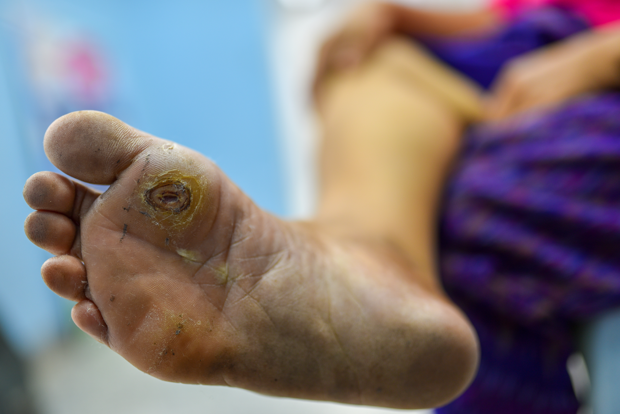
She doesn't recall which wound started first but does state that they started as painful purple areas that looked irregular and patchy. Soon afterward, she also developed painful bumps, and then everything seemed to just "open up." On examination, the new wounds all have a varying amount of soft to firm yellow slough at their bases with firm and dry brown eschar at the edges. The periwound skin has an erythematous border. You apply topical lidocaine to these wounds, and once the area is numb, you gently start to debride the wounds of their sloughy base and eschar. You are unable to remove all the slough, so you order an enzymatic debrider, which is to be used daily. You ask her to come back to the clinic in one week. Over the course of the week, you find yourself thinking about this patient and her strange new wounds.
Something doesn't sit right with you. The heel and hip wounds do seem to be related to pressure, but the calf and thigh wounds don't. The presentation seems odd as well. You recall from your education that pressure ulcers typically start as reddened areas that may blister and open. The patient described her skin as appearing purplish red and "lacy," which is more consistent with livedo reticularis (mottled purple skin) and doesn't typically form in relation to pressure. When the patient returns to the clinic the next week, you are disheartened to find that the wounds have increased in size and the slough at the base hasn't improved, despite the use of the enzymatic debrider. At this time, you pause and ask yourself what you're missing. You go into her chart again and look at the bloodwork she had while she was an inpatient.
Her kidney function remained poor, her white blood cell count was elevated, although it trended downward throughout her stay, and her albumin was on the low side. Her blood glucose levels ranged in the 100s to 200s, and her calcium and phosphorus levels were slightly elevated. Nothing strikes you as particularly off for her. This is another case of a "wound of the weird."
Pathogenesis and Risk Factors of Calciphylaxis
As you may have guessed by the title of this blog post, the wounds described in this hypothetical case are caused by calciphylaxis. Essentially, what can happen is that tiny occlusions form in the blood vessels of the subcutaneous fat and dermis and cause cutaneous ischemia and eventually death of the tissue. The occlusions are caused by calcification within the walls of the blood vessels.1
Think of this as a "heart attack of the skin." This can be a complication in patients with end-stage renal disease who are at the point of dialysis and when calcium and phosphate levels may become altered. This is known as uremic calciphylaxis, which occurs in most cases. The most common risk factor for developing calciphylaxis is renal failure, but other conditions that may result in calciphylaxis are anything that may cause the serum calcium or phosphate levels to fluctuate, such as thyroid disorders. Vitamin K deficiency may also lead to calcification of the blood vessels. Comorbid conditions and demographic factors that may put a patient at risk include the following1:
- End-stage renal disease requiring dialysis (especially if for more than six to seven years)
- Female sex
- Rapid weight loss
- Obesity
- Chronic illnesses: diabetes mellitus, liver disease, thyroid disease, autoimmune disease (lupus, temporal arteritis, rheumatoid arthritis), hypercoagulable conditions
- Altered laboratory results: hypercalcemia, hypoalbuminemia
Medications that may lead to increased risk are as follows2:
- Calcium supplements
- Calcium-based phosphate binders
- Active vitamin D
- Warfarin (Coumadin)
- Corticosteroids
- Iron therapy
- Trauma related to subcutaneous insulin or heparin injections
Diagnosis of Calciphylaxis
Diagnosis can be made based on clinical symptoms, but patients may benefit from further testing and laboratory work to evaluate for an underlying condition that may have contributed to the development of a calciphylaxis wound. Testing includes the following:
- Biopsy: look for calcification, microthrombosis, and fibrointimal hyperplasia of dermal or subcutaneous vessels2
- Laboratory tests to assess for risk factors2:
- Blood urea nitrogen-to-creatinine ratio and glomerular filtration rate
- Serum calcium, phosphorus, alkaline phosphatase, parathyroid hormone, 25-hydroxyvitamin D
- Liver testing: transaminase, alkaline phosphatase, and albumin
- Evaluation for infection: complete blood count with differential
- Coagulation evaluation: partial thromboplastin time
Clinical Manifestations
Clinical features include the following:
- Painful ulcers
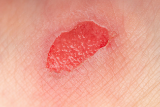
- Lacy pattern (livedo reticularis), which evolves to larger plaques and eventually black eschar
- Wound on areas with more subcutaneous fat, likely on the trunk and lower extremities3
- High mortality rate, with sepsis the leading cause of death1
Calciphylaxis wounds often start as small plaques that conjoin to form a larger patch of skin discoloration. It may have a lacy or "net-like" pattern that becomes purple or red and is very tender as a result of vascular ischemia. It may be confused with cellulitis at this stage. As the ischemia progresses, the plaques degrade into ulcers, which most commonly occur along sites of higher adiposity: the thighs and abdomen. In our hypothetical case, it is likely that the patient may have had several different types of wounds: the heel wound was probably pressure related, but the calf and thigh wounds are more indicative of calciphylaxis. Like most cases, this is certainly not cut and dry.
Treatment of Calciphylaxis
Treatment of calciphylaxis wounds can be difficult. Sadly, the annual mortality rate in patients who develop calciphylaxis is as high as 80%. Wound care should include surgical or manual debridement of devitalized tissue, hyperbaric oxygen therapy, and proper moisture balance ensured with appropriate dressings.
The overall goals of management should include excellent communication with the patient's entire health care team to ensure best practice management of the underlying disease that caused the vascular calcification. This should include ensuring proper calcium and phosphate levels, dietary support, and medication management. Sodium thiosulfate2 has been shown to be successful for decalcifying vessel walls, and it may be given intravenously during the last hour of a hemodialysis session. It may also be administered by direct injection around the wound and in the center of the ulcer. Vitamin K may also help to promote vascular decalcification.4 Treatment includes the following:
- Wound care with appropriate dressings, chemical debriding agents, and surgical debridement if needed
- Hyperbaric oxygen therapy if wounds are not improving
- Pain management as needed; pain can be severe, patient may need narcotic analgesics with an excellent pain plan
- Sodium thiosulfate: intravenous doses ranging from 12.5g to 25g in last 30 minutes of each hemodialysis session three times a week; may also provide intralesional sodium thiosulfate2
- Management of underlying chronic disease with multidisciplinary support, nutrition management, and management of other risk factors and medications
Conclusion
Once the appropriate interventions have been made, frequent follow-up is essential to ensure that the therapy is working and the wounds are improving. Calciphylaxis is an uncommon condition but one that is treatable. With the proper sleuthing and the right care, these wounds can be healed. Understanding that these wounds are like "a heart attack of the skin" can guide you in the right direction and help put your patient on the path to recovery.
References
1. Chang JJ. Calciphylaxis: diagnosis, pathogenesis, and treatment. Adv Skin Wound Care. 2019;32(5):205-215. doi: 10.1097/01.ASW.0000554443.14002.13. https://journals.lww.com/aswcjournal/Fulltext/2019/05000/Calciphylaxis_…. Accessed March 31, 2020.
2. Nigwekar SU, Kroshinsky D, Nazarian RM, et al. Calciphylaxis: risk factors, diagnosis, and treatment. Am J Kidney Dis. 2015;66(1):133-146. doi:10.1053/j.ajkd.2015.01.034. https://www.ncbi.nlm.nih.gov/pmc/articles/PMC4696752/. Accessed March 31, 2020.
3. Baby D, Upadhyay M, Joseph MD, et al. Calciphylaxis and its diagnosis: A review. J Family Med Prim Care. 2019;8(9):2763-2767. doi:10.4103/jfmpc.jfmpc_588_19. https://www.ncbi.nlm.nih.gov/pmc/articles/PMC6820424/. Accessed March 31, 2020.
4. Bhambri A, Del Rosso JQ. Calciphylaxis: a review. J Clin Aesthet Dermatol. 2008;1(2):38-41. https://www.ncbi.nlm.nih.gov/pmc/articles/PMC2989826/. Accessed March 31, 2020.
About the Author
Becky received her BSN from the University of Vermont where, along with a love of nursing, she picked up a love of hiking and cross-country skiing. She moved to Massachusetts and started to work as a med-surg nurse at a busy Boston hospital. There, she found that she loved mentoring new nurses and returned to school to earn her MSN as an acute care clinical nurse specialist from the University of Massachusetts, Boston. She followed her love of teaching into the acute, sub-acute and university settings, but she found that she missed working directly with patients. She returned to school and earned her Post-Master's Family Nurse Practitioner Certification from Rivier University. It was shortly after this that Becky discovered her love for wound care. She worked part time in wound care and part time in family care while she earned her WCC certification. After several years, Becky decided to take her practice to the next level by opening her own LLC and is currently seeing patients for wound care and regenerative medicine. Becky's philosophy of “Never stop learning” has guided her in her practice and life. Her very supportive husband and daughter are her key inspirations to keep growing and trying new things. In her spare time, Becky loves traveling with her family, going on long walks with her dog, Echo, and reading historical and science fiction.
The views and opinions expressed in this content are solely those of the contributor, and do not represent the views of WoundSource, HMP Global, its affiliates, or subsidiary companies.