Pedicure-Related Injuries, Part 2: Diagnostic Strategies, Treatment Protocols, and Prevention
May 5, 2026
Keywords
Categories
Pedicure-related injuries can present across a wide clinical spectrum, from mild periungual conditions to severe infections such as cellulitis, osteomyelitis, and necrotizing soft tissue disease. A structured diagnostic approach, timely intervention, and risk-based prevention strategies are essential for wound care practitioners to optimize outcomes and reduce complications.
Key takeaways
-
Recent pedicure exposure should be considered when evaluating new foot or ankle lesions, particularly in high-risk patients, as presentations may range from minor irritation to limb-threatening infection.
-
Thorough history, vascular and neurologic assessment, appropriate cultures (including for atypical organisms), and imaging are critical to accurately diagnose and guide management.
-
Prompt, severity-based management—ranging from local wound care to surgical intervention and broad-spectrum antibiotics—combined with patient education can prevent progression and improve outcomes.
Clinical presentations relevant to wound care
For wound care practitioners, recent pedicure exposure should be considered in the differential diagnosis when evaluating new‑onset toe, foot, or ankle lesions, particularly when onset closely follows a salon visit. Clinical manifestations span a continuum from localized periungual disease to extensive soft tissue involvement. Milder presentations may include acute and chronic paronychia, onycholysis, irritant or allergic contact dermatitis related to nail products, and small partial‑thickness wounds at sites of cuticle manipulation or callus reduction. While many of these lesions can be managed with local wound care, judicious debridement, and topical or short‑course systemic therapy, they warrant close follow‑up in high‑risk patients because of the potential for rapid deterioration. 1-4
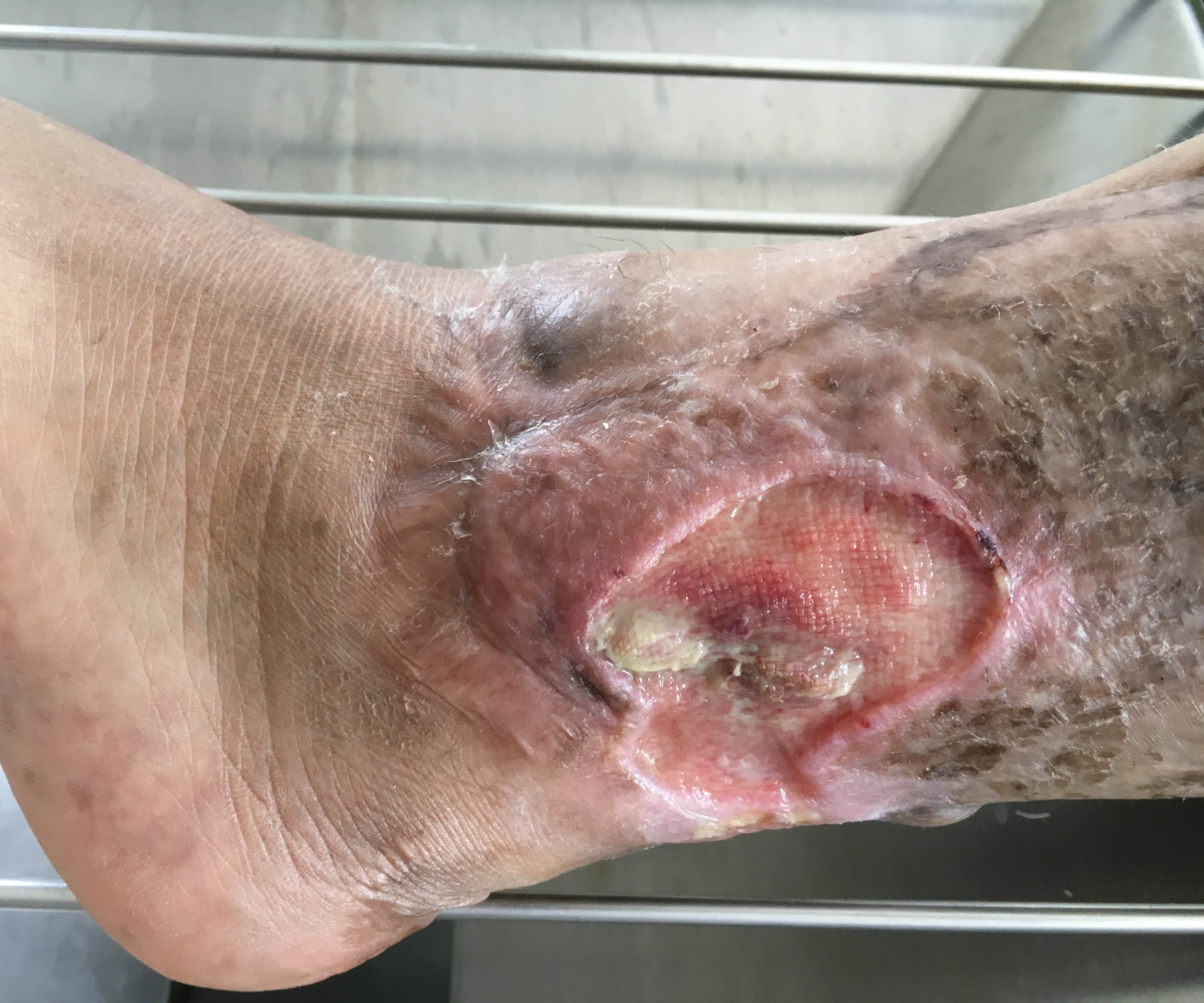
More substantial complications include cellulitis extending beyond the initial portal of entry, deep space abscesses, and traumatic wounds resulting from overly aggressive debridement or inappropriate use of sharp instruments. In persons with diabetes, peripheral arterial disease, or immunosuppression, such lesions may progress to osteomyelitis, necrotizing soft tissue infection, or chronic nonhealing ulcers requiring advanced therapies and, in some cases, amputation. Early recognition of a pedicure‑related mechanism, thorough vascular and neurologic assessment, appropriate microbiologic workup (including consideration of rapidly growing mycobacteria when standard cultures are negative), and timely referral for surgical consultation when indicated are therefore essential components of optimal management. 3-6
Diagnostic approach
A structured diagnostic strategy is essential when evaluating suspected pedicure-associated lower extremity complications. Comprehensive history-taking should specifically address the timing and nature of recent pedicure exposure, including the interval between the salon visit and onset of symptoms, details of the procedures performed, and patient observations regarding salon hygiene and instrument reprocessing practices, along with comorbid conditions that may impair healing such as diabetes, peripheral arterial disease, or immunosuppression. Physical examination should incorporate vascular assessment with palpation of pedal pulses and capillary refill, neurologic testing in at-risk patients (particularly those with diabetes and suspected neuropathy), and careful delineation of local and proximal infection spread, including inspection for lymphangitic streaking and regional lymphadenopathy. 7,8
Laboratory evaluation is recommended for moderate to severe presentations and should include complete blood count, inflammatory markers such as C-reactive protein and erythrocyte sedimentation rate, serum glucose, and blood cultures when systemic toxicity or sepsis is suspected. Wound sampling for microbiologic analysis ought to be performed prior to initiation of antimicrobial therapy, with deep tissue or aspirate specimens preferred over superficial swabs, and should encompass routine aerobic and anaerobic bacterial cultures as well as targeted mycobacterial studies when salon-associated exposure or atypical clinical patterns raise concern for rapidly growing nontuberculous mycobacteria. 7,8
Imaging plays a central role in assessing deep tissue involvement and excluding foreign bodies or underlying osteomyelitis. Plain radiography can identify subcutaneous gas, radiopaque foreign material, and bony changes compatible with chronic infection, whereas magnetic resonance imaging offers superior characterization of soft tissue planes, abscess formation, fascial involvement, and early bone marrow abnormalities suggestive of osteomyelitis. In patients with suspected arterial insufficiency, noninvasive vascular testing such as ankle-brachial index measurement, toe pressures, or duplex arterial ultrasonography should be pursued to guide both wound-healing prognosis and potential revascularization strategies. 7,8
Treatment protocol
Management should be individualized according to severity, with an emphasis on prompt, aggressive therapy to avert progression to limb- or life-threatening infection. Mild, localized lesions without systemic features may be managed on an outpatient basis with meticulous local wound care, including gentle cleansing with sterile saline, appropriate topical antimicrobial or antiseptic agents, edema control and limb elevation when indicated, and close surveillance for early signs of deterioration; concurrent discontinuation of irritant exposures and focused patient education about warning symptoms that warrant urgent reassessment are integral to this approach. 2,7,8
Moderate infections, characterized by more extensive cellulitis or early systemic manifestations, typically warrant empiric oral antimicrobial therapy directed against likely pathogens, including methicillin-resistant Staphylococcus aureus, streptococci, and selected Gram-negative organisms, pending culture data. Reasonable initial regimens may pair trimethoprim/sulfamethoxazole or doxycycline with a beta-lactam or fluoroquinolone to broaden coverage, with subsequent de-escalation or adjustment based on microbiologic results and susceptibility profiles; collections suggestive of abscess should be promptly incised and drained with appropriate submission of material for culture and histopathology. 2,7,8
Severe infections, including those with systemic toxicity, rapidly progressive soft tissue involvement, suspected necrotizing infection, or concomitant osteomyelitis, necessitate hospital admission, early surgical consultation, and parenteral broad-spectrum antibiotics, such as vancomycin in combination with piperacillin/tazobactam or a carbapenem, tailored to local resistance patterns and refined when culture results become available. Suspected or confirmed rapidly growing nontuberculous mycobacterial infections require prolonged, multidrug regimens—often incorporating macrolides (for example, clarithromycin), rifamycins, and additional agents based on species identification and susceptibility testing—in conjunction with surgical debridement when indicated. Adjunctive measures, including optimization of glycemic control in patients with diabetes, revascularization procedures for critical limb ischemia, use of negative pressure wound therapy for complex or highly exudative wounds, and consideration of hyperbaric oxygen therapy in selected necrotizing or refractory infections, may further improve outcomes in carefully chosen cases. Multidisciplinary collaboration involving wound care specialists, infectious diseases consultants, vascular and plastic surgeons, and, when appropriate, endocrinology or diabetes care teams is strongly recommended for complex presentations. 1-8
Prevention and patient education
Wound care clinicians occupy a pivotal position in prevention through risk stratification and targeted counseling. Patients with diabetes, significant peripheral arterial disease, prior foot ulceration, or immunosuppression should be advised to avoid commercial pedicures and instead obtain foot and nail care in medical settings where infection prevention and instrumentation standards are closely regulated.9,10 For lower-risk individuals who choose to continue salon-based services, counseling should emphasize selection of establishments with visible sterilization practices and current licensure, avoidance of razors or blades for callus reduction, refusal of cuticle cutting, preference for non–whirlpool basins or single-use liners, and use of personally owned, properly cleaned instruments when feasible; any breach in skin integrity during or shortly after a pedicure should prompt early clinical evaluation.3,5,9,10
Conclusion
Pedicure-related lower extremity complications remain an underrecognized cause of substantial morbidity and, in vulnerable patients, can progress to major limb loss. For wound care practitioners, maintaining a high index of suspicion through routine inquiry about recent salon exposures, structured diagnostic workup, and timely escalation to appropriately aggressive local and systemic therapy is critical to limiting progression and improving outcomes. In parallel, wound clinicians are uniquely positioned to deliver targeted patient education and risk-based counseling—particularly for individuals with diabetes, vascular disease, or immunosuppression—which represents a key strategy for primary prevention of these largely avoidable injuries.
References
1. Vugia DJ, Jang Y, Zizek C, Ely J, Winthrop KL, Desmond E. Mycobacteria in nail salon whirlpool footbaths, California. Emerg Infect Dis. 2005;11(4):616-618. doi:10.3201/eid1104.040936
2. Trevino EA, Weissfeld AS. Infections in nail salons. Clinical Microbiology Newsletter. 2008;30(2):9-11. doi:10.1016/j.clinmicnews.2008.01.001
3. Stout JE, Gadkowski LB, Rath S, Alspaugh JA, Miller MB, Cox GM. Pedicure-associated rapidly growing mycobacterial infection: an endemic disease. Clin Infect Dis. 2011;53(8):787-792. doi:10.1093/cid/cir539
4. Barn P, Chen T. A narrative review of infections associated with personal service establishments Part I: aesthetics. Environmental Health Review. 2012;55(1):9-26. doi:10.5864/d2011-002
5. Winthrop KL, Abrams M, Yakrus M, et al. An outbreak of mycobacterial furunculosis associated with footbaths at a nail salon. N Engl J Med. 2002;346(18):1366-1371. doi:10.1056/NEJMoa012643
6. ElSayed NA, Aleppo G, Aroda VR, et al. 2. Classification and Diagnosis of Diabetes: Standards of Care in Diabetes-2023. Diabetes Care. 2023;46(Suppl 1):S19-S40. doi:10.2337/dc23-S002
7. Cortes-Penfield NW, Armstrong DG, Brennan MB, et al. Evaluation and Management of Diabetes-related Foot Infections. Clin Infect Dis. 2023;77(3):e1-e13. doi:10.1093/cid/ciad255
8. Aldana PC, Cartron AM, Khachemoune A. Reappraising Diabetic Foot Ulcers: A Focus on Mechanisms of Ulceration and Clinical Evaluation. Int J Low Extrem Wounds. 2022;21(3):294-302. doi:10.1177/1534734620944514
9. Ju HH, Ottosen M, Alford J, Jularbal J, Johnson C. Enhancing foot care education and support strategies in adults with type 2 diabetes. J Am Assoc Nurse Pract. 2024;36(6):334-341. Published 2024 Jun 1. doi:10.1097/JXX.0000000000000998
10. Javaid K, Mistry S, Tchack M, Musolff N, Rafiq B, Rao B. Dermatologic Conditions Associated With Various Types of Popular Nail Cosmetics: A Systematic Review of Existing Literature and Future Recommendations. J Cosmet Dermatol. 2025;24(10):e70519. doi:10.1111/jocd.70519
Dr. Nirenberg is a clinical and forensic podiatrist, who has been in practice of over 33 years. He has assisted in educating cosmetologists on pedicure-related issues.
The views and opinions expressed in this content are solely those of the contributor, and do not represent the views of WoundSource, HMP Global, its affiliates, or subsidiary companies.