Scarring in Diverse Populations
Practice Accelerator
May 2, 2023
May 2, 2023
Keywords
Categories
Skin is considered the body's largest organ, and any damage can often lead to scarring. Although the pathology and physiology of what leads to scarring are not entirely understood, some details are concrete, including the series of processes that occur after the skin is damaged. These processes will be discussed in detail below, as well as the different types of scars. The appearance of these scars, as a result of these processes, may also present differently on various skin tones and types.
What are Scars?
A scar develops as part of the remodeling stage in the wound healing process and can commonly happen after surgery, skin trauma, or an infection.1 In more clinical terms, a scar results from a fibroblast replacement of regular skin tissue that didn't heal via regeneration.1 Instead, the body forms scar tissue to repair the wound through unidirectional collagen clumps instead of uniform collegian layers.1 Some scars may fade completely, and others may leave a mark.2 The type of wound and the length of time between occurrence and healing will often determine the scar’s depth, scope, and appearance.1 Specific factors that could contribute to a scar forming include “infections, retention of foreign bodies, and prolonged healing beyond 2-3 weeks.”1
How Do Scars Form?
This process is complex. Immediately after a wound occurs, the body attempts to repair itself through a variety of wound healing stages3,4:
- The first step is called hemostasis. The body attempts to clot the wound by sending platelets to the source of the injury. This process happens immediately after the wound occurs.
- The second step is inflammation. White blood cells attempt to fight off bacteria through phagocytosis. This phase often causes the skin to appear red, discolored, and even swollen.3 This phase should last anywhere from 1 to 4 days.
- The third step is proliferation. The skin creates new cells, causing the formed scab to shrink. After this process, the scab will be replaced with new skin cells. This phase can take anywhere from 4-21 days.
- Maturation, also known as remodeling, is the last step in wound healing, where new skin, including scarring, is visible. The skin can be in this phase post-injury for up to 2 years.
Other factors can affect scar formation, such as wound tension, infection, hypoxia, pregnancy, puberty, age, and cellular and genetic factors.5
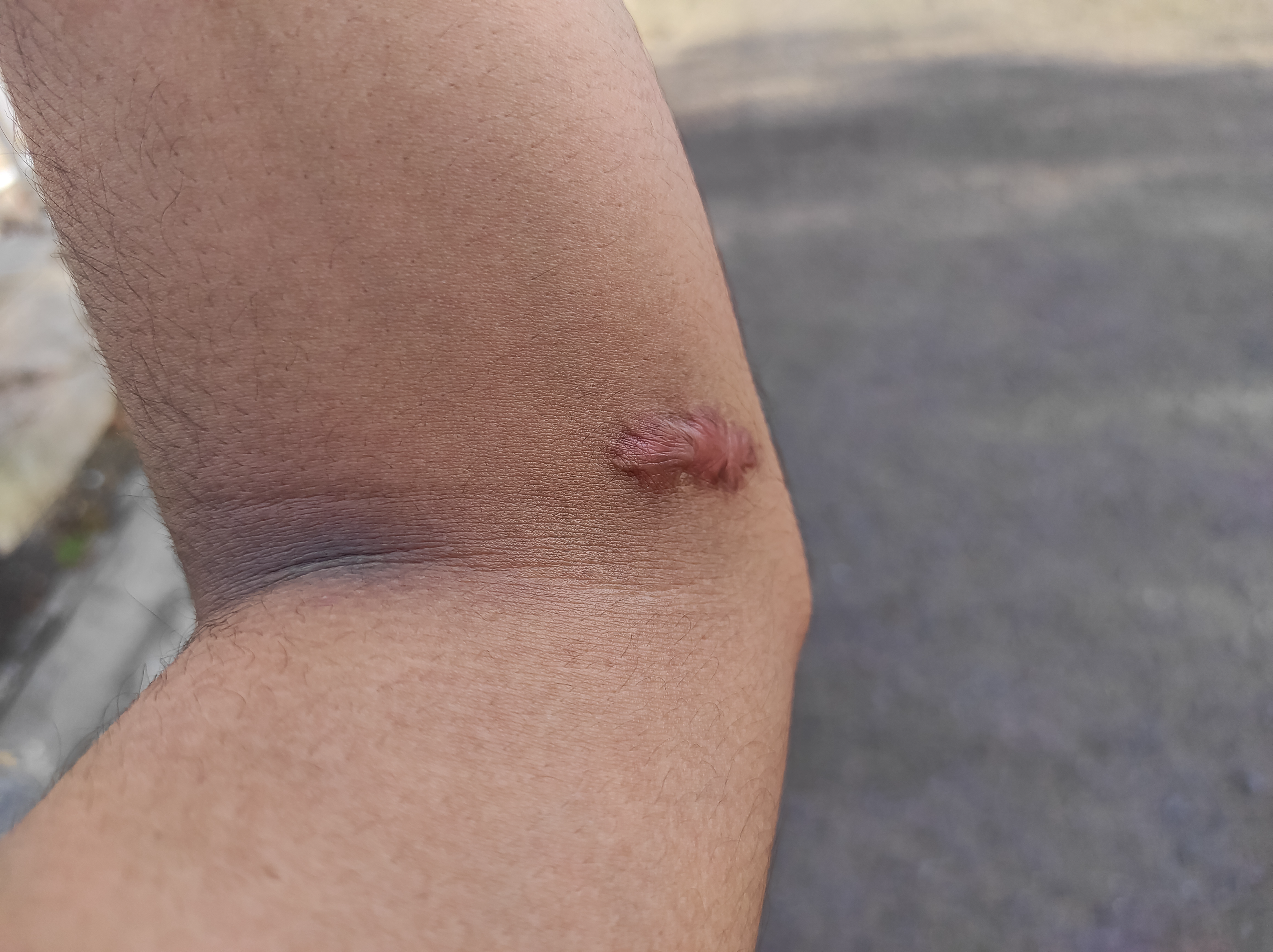
Different Types of Scars and Presentation in Skin Colors
A few types of scars have different presentations depending on skin tone and scar type. Keloid scars can appear dark or red and are thick and round. Unlike other types of scarring, it will appear and extend outside of the place where there was a wound.6 After a wound has healed, these scars result when the body produces too much collagen. As a result, they can often be painful. Keloids will develop anywhere and are challenging to treat.7-9 Hypertrophic scars appear raised and red.9 They may appear similar to keloid scar tissue, but hypertrophic scars are contained within the wound area, unlike a keloid scar.9 Like keloid scars, hypertrophic scars are more present in skin of color.9 Both keloid and hypertrophic scars tend to develop in places on the body where the tension of the skin is very tight. The presentation of these scars may look different on various skin tones, particularly in patients with skin of color.7 Although people of any skin color can get keloid or hypertrophic scarring, it is more prevalent in those with skin that is more pigmented. According to research that Tchero reported in their 2020 study “Management of Scars in Skin of Color,” 6-16% of African-Americans, Asians, and Hispanics developed keloids.7 Researchers have found that the greater the presence of melanin in the body, the more likely one is to experience the development of keloids.8 When skin is injured, in addition to other cellular processes, the body produces cells called melanocytes, which, put simply, means that there is an increase of melanin in the area.8 Additionally, as a wound goes through the stages of healing, these melanocytes may be destroyed in the process. As the skin heals, these cells return in varying degrees, which is why some scars may appear lighter or darker than the surrounding skin.10
Treatment
Both forms of scarring, keloid and hypertrophic, may create discomfort and diminish patient’s self-perceptions.11 A study looked at how African Americans and white patients' perception of their scars impacted how they viewed their “appearance, psychosocial health, and career,” for instance.11 The study found that African Americans were more likely than white patients to have a lower self-perception. These findings point to the larger need for clinicians to consider a patient’s individual needs when creating a treatment plan.11 Several treatments for scar minimization exist, including the following12:
- Steroid injections
- Silicone sheets
- Cryotherapy
- Excision and laser surgery
One in particular, nonablative fractional laser resurfacing, has been found to be effective in treating scarring in patients with skin of color.12 Nonablative fractional lasers target water instead of melanin in the skin and, as a result, are safer for use on darker skin. However, more information and research about its use are still taking place. Although fractional lasers are safer than traditional lasers for those with darker skin, side effects are still present, including edema, hyperpigmentation, and transient erythema.12
Conclusion
There are various treatments for scars; however, there isn't a way to remove them completely. Some treatments have the potential to worsen a scar.11 Understanding how scars present is essential for managing them in patients of varying skin tones. Giving patients a proper treatment plan can maximize the opportunity for a positive outcome.
References
- Al-Shaqsi S, Al-Bulushi T. Cutaneous Scar Prevention and Management: Overview of current therapies. Sultan Qaboos Univ Med J. 2016;16(1):e3-e8. doi:10.18295/squmj.2016.16.01.002
- American Society for Dermatological Surgery. Scars. Accessed March 29, 2023. https://www.asds.net/skin-experts/skin-conditions/scars
- Hultman S. Everyday Cuts and Scrapes: How to Prevent Scarring. Johns Hopkins Medicine. Accessed March 29, 2023. https://www.hopkinsmedicine.org/health/wellness-and-prevention/everyday…
- Nova Scotia Health. Skin and Wound Care. Updated March 21, 2023. Accessed March 29, 2023. https://library.nshealth.ca/WoundCare/HealingBasics
- Aydoğmuş S, Kelekçi KH, Şengül M, et al. Factors affecting the development of scar formation in abdominal surgery performed for gynecologic and obstetric conditions. Turkderm-Turk Arch Dermatol Venereology. 2017;51:12-7. doi: 10.4274/turkderm.58751
- Stanford Medicine. Keloid Scars. Accessed March 29, 2023. https://stanfordhealthcare.org/medical-treatments/s/scar-revision-surge…
- Tchero H. Management of Scars in Skin of Color. In: Téot L, Mustoe TA, Middelkoop E, Gauglitz, GG (eds). Textbook on Scar Management. Springer, Cham. 2020. https://doi.org/10.1007/978-3-030-44766-3_43
- Ludmann P. Keloid Scars: Causes. American Academy of Dermatology Association. Updated August 8, 2022. Accessed March 29, 2023. https://www.aad.org/public/diseases/a-z/keloids-causes
- Xue M, Jackson CJ. Extracellular Matrix Reorganization During Wound Healing and Its Impact on Abnormal Scarring. Adv Wound Care. 2015;4(3):119-136. doi:10.1089/wound.2013.0485
- McGrory C. Reducing the impact of hypertrophic scarring. Wounds UK. 2013;9(3):18-22. https://www.wounds-uk.com/resources/details/reducing-impact-hypertrophi…
- Garg SP, Hassan AM, Patel A, et al. Scar Perception: A Comparison of African American and White Self-identified Patients. Plast Reconst Surg. 2022;10(5):p e4345. DOI: 10.1097/GOX.0000000000004345
- Kaushik SB, Alexis AF. Nonablative Fractional Laser Resurfacing in Skin of Color: Evidence-based Review. J Clin Aesthet Dermatol. 2017;10(6):51-67.
The views and opinions expressed in this content are solely those of the contributor, and do not represent the views of WoundSource, HMP Global, its affiliates, or subsidiary companies.
More from this Author
// fixed missing link variable.
// fixed missing link variable.
// fixed missing link variable.


