Conducting a Biomechanical Exam As Part of Pressure Injury Prevention
Practice Accelerator
November 1, 2023
November 1, 2023
Keywords
Categories
Introduction
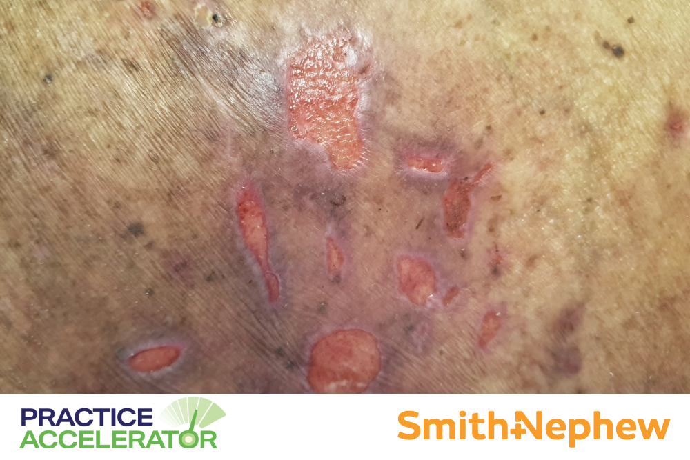
Pressure injuries (PIs) often result from sustained pressure and shear on skin and local tissue. As a result, patient position, posture, and load bearing are integral factors to consider to alleviate such forces. A better understanding of the biomechanics of these factors may assist clinicians and scientists in identifying and addressing biomechanical risk factors for pressure injury. As a result, research and design improvement of medical equipment could contribute to preventing device-related pressure injuries DRPIs,1 and clinicians may have better insights into high-risk areas for skin injury.
Why Do Pressure Injuries Occur from a Biomechanical Perspective?
Epidemiologically and based on anatomic and biomechanical function, PIs in adults are often at the heels, sacrum, or ischial tuberosities.1 These locations of injury are in part due to the mechanics associated with prolonged supine or sitting positions. When a patient is placed in these positions for long periods, these bony prominences take on more pressure. Conversely, thinking about the body mechanics of a neonate, looking at body habitus and function as it relates to typical positioning, the head is more prominent, and therefore the occiput is more typically involved in PI development.1 Other mechanical factors that contribute to pressure injury risk include:
- Mobility
- Contracture
- Spasticity
Spasticity is a patient's ability to move within that position to naturally offload high-risk spots. Can they shift their weight from side to side, or can they roll over on their own? If not, how can clinicians assist effectively, and/or what devices are available in the facility that may support offloading?
What Recent Literature Points Out
Computer-aided analysis and modeling have been studied concerning the impact of biomechanics on pressure injuries. Gefen found that computerized biomechanical modeling showed that atypical foot anatomy, specifically a sharp posterior calcaneus and thin, soft tissue padding, predicted risk for pressure injury.2 Combining this factor with potential loss of mobility (less ankle range of motion) and a patient’s natural heel position (which part of the heel contacts the bed or chair), one can logically see how biomechanics can provide valuable information in preserving skin integrity.
Biomechanical Assessment
Experts also add that glycosylation of tissues and tissue flexibility changes related to diabetes or other conditions can play a role in altering a patient’s biomechanics, thus placing them at risk for skin breakdown and pressure injury.3 Thought leaders advocate for a biomechanical examination starting with a wound's initial presentation, and in high-risk patients, even before a wound develops.
"Their biomechanical history is extremely important to look at: the tissue flexibility, the presence of hallux limitus, posterior tibial problems, bunions, hammertoes, all the malformations that the foot takes on over a lifetime," said James McGuire, DPM, PT, CPed, CWS, FAPWCA, and a Professor in the Department of Podiatric Medicine and Biomechanics at Temple University School of Podiatric Medicine.3
How Much Do You Know About Pressure Injury Prevention? Take our quiz to find out! Click here.
For the lower extremity, a full assessment of gait, stance, range of motion, and deformities, among other factors, is important to identify how a patient functions and how these factors relate to incurring pressure, friction, or shear forces. As with other areas of the body, this pressure can come from sources such as:
- Clothing (shoe or sock wear)
- Bedding
- Walking surface
- Resting surfaces (chair or bed)
- Medical devices (casts, walking boots, or prosthetics)
A biomechanics specialist related to the area of concern could be a wise referral for a patient at risk. This referral could include a podiatrist for lower extremity concerns or a physical therapist.
Perioperative Repositioning
In the perioperative arena, positioning is a vital component of a procedure's success. However, the patient's body mechanics, or limitations thereof, can pose challenges and increased risk for pressure injury. An operating room does not offer the same forms of positioning adaptations as other clinical settings.
An optimal perioperative position for a patient may be a compromise arrived upon by weighing the needs for adequate surgical access and visualization with the resultant pressures exerted by the operating table, anesthesia, other medical devices, and safety equipment utilized.4 The most commonly used position is supine, which has multiple variations.
Considering the biomechanical impact of these position changes may shed light on pressure injury risk. For instance, if a gel roll is used to hyperextend the neck, allowing better access to the chest, the posterior neck is at higher risk for PI.4 Each mechanical positioning schematic carries different functional and pressure-related challenges. However, proper preoperative evaluation techniques and use of appropriate positioning devices are just a few ways that clinicians can use biomechanics to alleviate these challenges. Gefen and team performed a scoping review that stressed the multi-pronged biomechanical approach to protecting tissue in the operating room.4
Conclusion
Biomechanics as it relates to wound development, including pressure injury, is an area in need of continued research. Most of the current literature focuses on soft tissue mechanics from the point of view of how these structures function under tension or stretch. Whereas, in the wound care world, clinicians need to know how these structures function under compression.5 However, by incorporating a biomechanical point of view into one's evaluation of patients at risk of pressure injury, clinicians may be able to discern new and helpful actionable information to work toward prevention.
References
- Levy A, Kopplin K, Gefen A. Device-related pressure ulcers from a biomechanical perspective. J Tissue Viability. 2017;26(1):57-68.
- Gefen A. The biomechanics of heel ulcers. J Tissue Viability. 2010;19(4):124-131.
- Suzuki K, McGuire J. Biomechanics in wound care. Podiatry Today Podcasts. Published May 1, 2023. Accessed October 25, 2023. https://www.hmpgloballearningnetwork.com/site/podiatry/podcasts/biomech….
- Gefen A, Creehan S, Black J. Critical biomechanical and clinical insights concerning tissue protection when positioning patients in the operating room: a scoping review. Int Wound J. 2020;17(5):1405-1423.
- Jan Y-K, Major MJ, Pu F, Sonenblum SE. Editorial: soft tissue biomechanics in wound healing and prevention. Front Bioeng Biotechnol. 2022:10. https://doi.org/10.3389/fbioe.2022.897860.
The views and opinions expressed in this content are solely those of the contributor, and do not represent the views of WoundSource, HMP Global, its affiliates, or subsidiary companies.
More from this Author
// fixed missing link variable.
// fixed missing link variable.
// fixed missing link variable.


