Getting to Know Granulation Tissue and What it Means for Wound Care
February 11, 2021
Keywords
Categories
As a wound care nurse practitioner, when I see granulation tissue start to form on a wound, I do a little happy dance. Granulation tissue is a sign that the wound is on its way past an often-stubborn inflammatory phase of healing and progressing into the building phase of proliferation. But what exactly is granulation tissue? And why does its presence indicate that the wound is healing? Let’s explore this a bit more.
What Does Granulation Tissue Look Like?
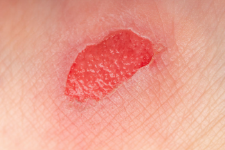
Granulation tissue often appears as red, bumpy tissue that is described as “cobblestone-like” in appearance. It is highly vascular, and this is what gives this tissue its characteristic appearance. It is often moist and may bleed easily with minimal trauma. You may see this tissue start to slowly fill in the wound in small, papular-like developments or in a more widespread pattern. Granulation tissue is the primary type of tissue that will fill in a wound that is healing by secondary intention. It is made up of macrophages, which help to remove debris and release cytokines. Cytokines help to activate fibroblasts, which will make collagen, trigger endothelialization, and help with the formation of new blood vessels, a process known as angiogenesis. We will also see granulocytes in granulation tissue, which are a type of white blood cell that can help fight infection.
What Does The Presence of Granulation Tissue Mean?
In short, observing granulation tissue in the bed of the wound means that the wound is progressing from the inflammatory phase of healing to the proliferative phase of healing. Several important cellular developments are occurring. Matrix metalloproteinases (MMPs), which are so helpful in removing damaged tissue and bacteria from the inflammatory phase, have started to allow the formation of new blood vessels at the wound bed. The number of MMPs is now starting to drop, which is a good thing because chronic MMPs can actually cause degradation of healthy proteins and growth factors and may delay healing. Cytokines, which are cells that are triggered by macrophages, are starting to increase in numbers and are telling the fibroblasts to get to work to start forming new tissue and blood vessels. You will also likely see a reduction in the four classic signs of inflammation: edema and erythema of the periwound, pain, and heat, which also indicate that the wound is progressing into the proliferative phase of healing.1,2
Types of Granulation Tissue
Hypotrophic Granulation Tissue
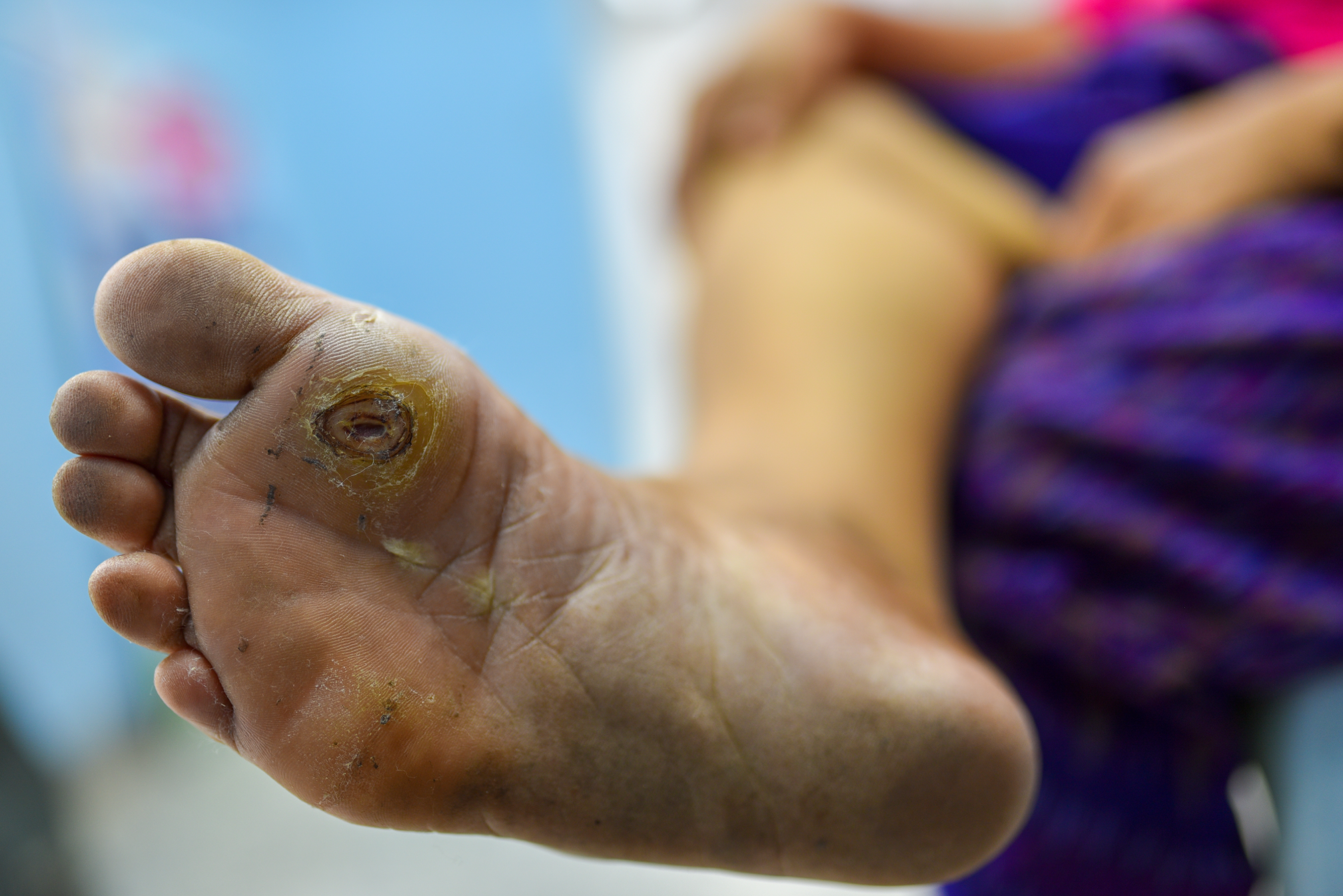
There are several variations of granulation tissue that you may encounter. You may find that the wound is filling in with new tissue; however, unlike the classic moist, beefy red tissue, it may appear smooth, pink, or even slightly pale. This is hypotrophic granulation tissue. I think of this as a wound that is desperately trying to heal, but something is standing in the way. It indicates poor perfusion and often is caused by pressure, poor circulation, trauma, or infection.1 If you observe this, it is important to assess for these factors because the wound is unlikely to improve until these issues are corrected. Make sure to offload any pressure, evaluate for potential trauma, and assess for and treat infection if present. This should help to alleviate hypotrophic granulation tissue and allow for healthier granulation tissue to develop.
Hypertrophic Granulation Tissue
Another type of granulation tissue that you will likely observe is hypertrophic granulation tissue. I think of this as granulation tissue growth on overdrive. It will still have that classic moist, beefy red appearance, but it will be raised above the surface of the wound. This will prevent the migration of epithelial cells across the center of the wound and will hinder healing. It is often a sign of excessive moisture or even infection, so make sure that you evaluate for this. After assessing for and treating these factors, some other interventions that you may consider are:
- Cauterization of the hypertrophic tissue with silver nitrate. This will effectively “beat back” the tissue and help control the overgrowth.
- Treatment with a daily topical steroid cream for one to two weeks. This can manage the overgrowth and allow for epithelial progression across the wound surface. Ensure that this cream is not used for more than 14 days because it can lead to excessive tissue atrophy.
- Utilization of a polyvinyl alcohol–gentian violet-methylene blue foam dressing. This has shown to be an effective dressing to manage hypertrophic granulation tissue.
You may be wondering whether there are any interventions that you can implement to help encourage the formation of granulation tissue. You can help encourage the proliferation of granulation tissue by:
- Managing exudate
- Selecting dressings to provide a moist, warm healing environment
- Removing any necrotic tissue, which may cause MMPs to linger in excessive amounts
- Ensuring that the patient is properly nourished and hydrated
- Assessing and treating any underlying comorbidities
Conclusion
With these factors in mind, once you do start to observe granulation tissue formation, it is important to ensure that the wound is protected. This is a good time to start applying a collagen dressing or, for deeper wounds, negative pressure wound therapy. Try to space the dressings out as appropriate to every other day or even several times a week to provide a constant warm, moist environment for healing. With these tips in mind, the granulation tissue can continue to fill in the wound bed and allow the wound to contract and close in. I hope that you, too, will celebrate a bit once you start to see the formation of granulation tissue because it is truly a beautiful thing.
References
1. Alhajj M, Bansal P, Goyal A. Physiology, Granulation Tissue. Treasure Island: Stat Pearls; 2020. https://www.ncbi.nlm.nih.gov/books/NBK554402/#:~:text=The%20granulation…. Accessed July 19, 2020.
2. Landén NX, Li D, Ståhle M. Transition from inflammation to proliferation: a critical step during wound healing. Cell Mol Life Sci. 2016;73(20):3861-3885. https://www.ncbi.nlm.nih.gov/pmc/articles/PMC5021733/. Accessed July 19, 2020.
About the Author
Becky received her BSN from the University of Vermont where, along with a love of nursing, she picked up a love of hiking and cross-country skiing. She moved to Massachusetts and started to work as a med-surg nurse at a busy Boston hospital. There, she found that she loved mentoring new nurses and returned to school to earn her MSN as an acute care clinical nurse specialist from the University of Massachusetts, Boston. She followed her love of teaching into the acute, sub-acute and university settings, but she found that she missed working directly with patients. She returned to school and earned her Post-Master's Family Nurse Practitioner Certification from Rivier University. It was shortly after this that Becky discovered her love for wound care. She worked part time in wound care and part time in family care while she earned her WCC certification. After several years, Becky decided to take her practice to the next level by opening her own LLC and is currently seeing patients for wound care and regenerative medicine. Becky's philosophy of “Never stop learning” has guided her in her practice and life. Her very supportive husband and daughter are her key inspirations to keep growing and trying new things. In her spare time, Becky loves traveling with her family, going on long walks with her dog, Echo, and reading historical and science fiction.
The views and opinions expressed in this content are solely those of the contributor, and do not represent the views of WoundSource, HMP Global, its affiliates, or subsidiary companies.
More from this Author
// fixed missing link variable.
// fixed missing link variable.
// fixed missing link variable.