Is It Really Infected? Recognizing Infection in Diabetic Foot Ulcers Based on the Evidence
February 25, 2022
Keywords
Categories
Background
Standards of care and evidence-based guidelines should lead our wound care practice to ensure the best possible outcomes for our patients. There are often prewritten algorithms or first- and second-line therapies, along with outlined treatment plans and guidelines established based on evidence. These guidelines can be adjusted to meet each patient’s specific needs.
Establishing Guidelines for Infection
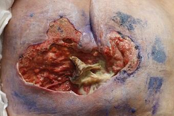
When determining whether a wound is infected, it is important to look at the big picture and to differentiate among infection, inflammation, contamination, and colonization. Additionally, it is important to rule out other etiologies (for example, dependent rubor versus true erythema of an extremity) and incorporate evidence-based guidelines into our treatment plan. The Wound, Ostomy and Continence Nurses (WOCN®) Society released updated guidance in November of 2021 related to management and treatment of patients with lower extremity wounds resulting from diabetes mellitus and/or neuropathic disease.1 The WOCN® Guideline for Management of Patients With Lower-Extremity Wounds Due to Diabetes Mellitus and/or Neuropathic Disease contains the evidence-based recommendations of a task force guided by levels of evidence and current standards of care.1 This short blog will focus on identifying and diagnosing infection in lower extremity wounds, based on the recommendations from this evidence-based guideline.
Assessment
Assessment should include a complete physical evaluation, including a head-to-toe physical examination. It is important to gather a medical, surgical, and family history to and look carefully at the patient’s current and past comorbid conditions. Gathering thorough background information and performing a current assessment are pertinent in accurately identifying current wound etiology and establishing an evidence-based treatment plan. An assessment for local and systemic signs or symptoms of infection should be conducted during this process.
Biofilm
If a wound fails to heal despite appropriate offloading and proper diagnostic and therapeutic interventions, biofilm may be the culprit. Biofilms are not visible to the naked eye but may be seen under a microscope. Clinical diagnosis may be made in the absence of definitive diagnostic options for biofilm management. Some clinical and assessment findings indicative of chronic biofilm may include:
- Necrotic tissue or the presence of slough in the wound bed
- Negative culture despite optimal sample
- Medical history of a biofilm-predisposing condition (e.g., implanted device)
- Prolonged inflammatory phase of healing
- Friable granulation tissue
- Presence of undermining within the wound
- No response to topical or systemic antimicrobial therapy
Patients with a severe diabetic foot infection should be referred to a specialist within 24-hours because of the high risk of complications and the presence of infection. If biofilm is suspected based on clinical findings or microscopic review, a tissue specimen may be helpful for culture (curette or biopsy) to determine the causative pathogens and their sensitivity to antibiotics or in clinically infected wounds. If tissue samples are not available, quantitative swabs using the Levine technique are a reasonable alternative. Wound cultures are performed to identify both aerobic and anaerobic bacteria and should be avoided when clinical indications are not clearly present.1
Imaging
The WOCN® 2021 guideline recommends obtaining serial, plain radiographs (x-rays) in patients with a new diabetic foot infection to examine the foot for foreign bodies, bone abnormalities or deformity, or soft tissue gas.1 Magnetic resonance imaging (MRI) should be used for patients when there is a concern for abscess or underlying bone infection (osteomyelitis). If MRI is unavailable or contraindicated, a leukocyte scan combined with a bone scan is an appropriate alternative.1
Osteomyelitis
In addition to a thorough clinical assessment, inflammatory biomarkers may be used to determine whether osteomyelitis is present. White blood cell (WBC) count, C-reactive protein (CRP), and erythrocyte sedimentation rate (ESR) are commonly reviewed laboratory results when looking for inflammation or infection. Other conditions that cause inflammation or an increase in these inflammatory markers should also be taken into consideration when reviewing the entire clinical picture. The WOCN® guideline recommends using a combination of plain radiographs, probe-to-bone testing, and laboratory studies as an initial workup to diagnose osteomyelitis.1 Radiographs have low sensitivity and specificity to confirm or exclude a diagnosis of osteomyelitis, but they can be helpful in ruling out other conditions. If the diagnosis is in doubt, MRI or a leukocyte scan combined with a bone scan is recommended. When making a definitive diagnosis of osteomyelitis, a sample of the bone (bone biopsy) is recommended to culture for microorganisms and histopathologic examination, to guide treatment.1
Conclusion
Diabetic foot ulcers can often be refractory or slow healing and predisposed to infection for a multitude of reasons. These wounds require an interprofessional approach and an effective, evidence-based management plan for optimal healing and prevention of recurrence. Infection and/or colonization should be looked at and ruled out or treated to achieve the best outcomes and healing potential for each individual patient. An evidence-based guideline is a great tool to use to ensure that we are following a standard of care and the most up-to-date best treatment practice.
Reference
- Wound, Ostomy and Continence Nurses Society. WOCN® Guideline for Management Of Patients With Lower-Extremity Wounds Due to Diabetes Mellitus and/or NEUROPATHIC DISEASE. Mt. Laurel, NJ: Wound, Ostomy and Continence Nurses Society; 2021.
About the Author
Holly is a Wound Care Coordinator at The Department of Veterans Affairs Medical Center in Cleveland, Ohio. She has a passion for education, teaching, and our Veterans. Holly has been practicing in wound care nursing for approximately six years. She has much experience with the long-term care population and chronic wounds as well as pressure injuries, diabetic ulcers, venous and arterial wounds, surgical wounds, radiation dermatitis, and wounds requiring advanced wound therapy for healing. Holly enjoys teaching new nurses about wound care and most importantly, pressure injury prevention. She enjoys working with each patient to come up with an individualized plan of care based on their needs and overall medical situation. She values the importance of taking an inter-professional approach with wound care and prevention overall, and involves each member of the healthcare team as much as possible. She also values the significance of the support of leadership within her facility and the overall impact of great teamwork for positive outcomes.
The views and opinions expressed in this content are solely those of the contributor, and do not represent the views of WoundSource, HMP Global, its affiliates, or subsidiary companies.
More from this Author
// fixed missing link variable.
// fixed missing link variable.
// fixed missing link variable.