Incontinence-Associated Dermatitis: Evidence-Based Practices
February 10, 2023
Keywords
Categories
Introduction
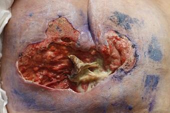
Moisture-associated skin damage (MASD) is sometimes accompanied by serous exudate, a denuded appearance of skin, or a secondary cutaneous infection. As the most common form of MASD, incontinence-associated dermatitis (IAD) is characterized by erythema and edema of the skin’s surface. IAD is a top-down injury, often presenting with inflammation, erosion, or denudation in the setting of fecal or urinary incontinence.1 It is a significant problem as it can result in the following complications for patients1:
- Infection
- Increased risk for pressure injuries
- Pain and discomfort
- Loss of independence and disruption in activities (sleep/wake cycle)
- Decreased quality of life (QoL) and depression
This condition is prevalent as it is often seen in acute care settings, long-term care, residential care and among community-dwelling individuals, or those living at home. Studies have shown that IAD is an independent risk factor for pressure injuries, placing those with fecal or urinary incontinence at higher risk for developing a pressure injury across care settings.1
What Causes Incontinence-Associated Dermatitis?
Fecal or urinary incontinence alone increases the risk for IAD. Overhydration (increased moisture) of the stratum corneum compromises the skin’s epidermal layer configuration. As a result, irritants from urine and stool penetrate this layer and cause inflammation and impair tensile strength.1 The frequency of incontinence also impacts the risk for IAD, along with the following1:
- Prior scarring or history of pressure injury
- Pre-existing comorbidities
- Immobility or compromised mobility
- Medications
- Nutritional status
- Inability to perform activities of daily living/cleansing skin independently
- Level of cognitive functioning
- Overall skin condition
Additionally, skin injury by friction, commonly defined as “resistance to motion in a parallel direction relative to the common boundary of 2 surfaces,"1 is a risk factor for worsening IAD in the moisture setting. It often presents as initial erythema or abrasion.
How to Assess IAD
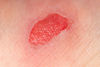
Clinical assessment may show initial erythema that ranges from pink to red. In patients with dark skin tones, IAD may present with more pale, yellow, or dark discoloration. Skin affected by IAD can appear as patchy discoloration with irregular borders and may present with vesicles, pustules, or papules. A red rash with pin-point satellite lesions may indicate a fungal infection (candidiasis) and is not uncommon.¹
Patients may also complain of burning, itching, tingling, or pain in the affected areas. Patients who experience IAD should have skin assessments at regular intervals to check for signs of worsening IAD or deterioration of skin condition. In addition, wound care professionals should ensure that appropriate prevention and treatment measures are in place. Management of IAD depends on clinical assessment, the severity of skin damage, and the type of incontinence (fecal, urinary, or mixed).1
Differential diagnoses most commonly include stage 2 pressure injuries, friction (superficial, top-down wounds), or shear (bottom up) injuries.1 It is vital to pay close attention to the anatomical location of skin injury along with environmental factors and other pre-existing comorbidities to diagnose accurately and formulate a comprehensive plan of care, addressing not only skin injury but all contributing factors.
How to Prevent and Manage IAD
The best treatment strategy is, of course, prevention, but as we know, IAD cannot always be prevented. It must be managed with containment, topical therapies, and lifestyle modification or assistive devices. The most effective way to prevent IAD from worsening is to eliminate or reduce incontinent episodes. This approach removes the primary factors like moisture, skin irritants, and friction contributing to skin injury.1 Methods to prevent IAD include1:
- Engaging patients in bowel and bladder retraining programs
- Attention to mobility status/promoting mobility
- Identifying and modifying (times, usage) of problematic medications in collaboration with providers and pharmacy
- Establishing a toileting schedule
- Setting care goals
Clinicians should involve an interdisciplinary team and treat not just the skin but additional causative/problematic factors within the environment and those impacting the patient. This approach will yield the best outcomes when preventing and managing IAD, a condition so significant across care settings.
Looking Towards the Future
As a condition that increases hospital stay and reduces quality of life, IAD is a formidable obstacle. A 2020 literature review from the Journal of Wound, Ostomy, and Continence Nursing found that, although there is extensive evidence on the factors that contribute to IAD development, there is a gap in evidence concerning its "underlying mechanisms."2 Areas of suggested research included the effect of urine on skin pH and the impact of digestive enzymes and bacteria present in stool on skin integrity.2
Currently, some of the pathophysiological components that contribute to IAD development are known. The presence and amount of ammonia in urine, especially malodorous urine, has been found to facilitate bacteria growth by affecting the pH of weakened skin. In addition, the digestive enzymes present in liquid stool macerate skin, expedite the release of cytokines, and decrease the skin's barrier function.3
A researcher from the 2020 literature review, joined by several other researchers, conducted a study in 2022 on the development of IAD in rats as a model for future, more effective prevention and management methods.3 Bacteria in combination with urine resulted in IAD, especially when skin was treated with sodium lauryl sulphate, which emulated recurrent surfactant use and irritated skin. Ultimately, the researchers found that bacteria are a major deciding factor in IAD development. The researchers recommended using pH balanced cleansers and similar products to preserve the skin's barrier function, which is often diminished across the aging population.3
Conclusion
Evidence-based practices are constantly evolving in wound care. For IAD, further research will aid in advancing prevention and management methods and, therefore, ensure better outcomes for patients. Meanwhile, wound care professionals can educate themselves on evidence-based best practices in an ever-evolving medical landscape.
References
- Thayer D, Nix D. Incontinence Associated Dermatitis. In: Ermer-Seltun J, Engberg S, ed. Wound, Ostomy, and Continence Nurses Society Core Curriculum: Continence Management. 2nd ed. Philadelphia, PA: Wolters Kluwer; 2021:364-381.
- Koudounas S, Bader DL, Voegeli D. Knowledge Gaps in Etiology and Pathophysiology of Incontinence-Associated Dermatitis: A Scoping Review. JWOCN. August 2020; 47(7): 388-395. doi: 10.1097/WON.0000000000000656
- Koudounas S, Minematsu T, Mugita Y, et al. Bacterial invasion into the epidermis of rats with sodium laurel sulphate-irritated skin increase damage and induces incontinence-associated dermatitis.J Inter Wound. August 2, 2022; 20(1): 191-200. https://doi.org/10.1111/iwj.13864
About the Author
Holly is a board certified gerontological nurse and advanced practice wound, ostomy, and continence nurse specialist at VA Northeast Ohio Healthcare System in Cleveland, Ohio. She has a passion for education, teaching, and our veterans. Holly has been practicing in WOC nursing for approximately ten years. She has much experience with the long-term care population and chronic wounds as well as pressure injuries, diabetic ulcers, venous and arterial wounds, surgical wounds, radiation dermatitis, and wounds requiring advanced wound therapy for healing. Holly enjoys teaching new nurses about wound care and, most importantly, pressure injury prevention. She enjoys working with each patient to come up with an individualized plan of care based on their needs and overall medical situation. She values the importance of taking an interprofessional approach with wound care and prevention overall, and involves each member of the health care team as much as possible. She also values the significance of the support of leadership within her facility and the overall impact of great teamwork for positive outcomes.
The views and opinions expressed in this content are solely those of the contributor, and do not represent the views of WoundSource, HMP Global, its affiliates, or subsidiary companies.
More from this Author
// fixed missing link variable.
// fixed missing link variable.
// fixed missing link variable.