How Inflammation Affects Wound Healing
Practice Accelerator
July 22, 2021
July 22, 2021
Keywords
Categories
An injury to the human body initiates a wound healing chain reaction that occurs in four sequential but overlapping phases: hemostasis, inflammatory, proliferative and maturation. This post focuses on the second (inflammatory) phase, which begins after blood flow stops (ie, hemostasis) and defender white blood cells, or leukocytes, migrate to the site of the injury — a process known as chemotaxis.1
Understanding the Inflammatory Stage of Wound Healing
The inflammatory stage typically lasts several days, but it can go on for much longer, making the wound chronic. Many cells and chemical reactions or signals keep the wound progressing in the inflammatory phase. Understanding these processes can jump-start a chronically stalled wound so that healing resumes. The clinician’s goals in the inflammatory phase are to limit further damage, close the wound, remove cellular debris and bacteria, and encourage cellular migration.1 Following hemostasis and chemotaxis, white blood cells and thrombocytes release more mediators and signaling cytokines, which accelerates the inflammatory process.
Several growth factors work in concert to promote collagen degradation, transform fibroblasts, grow new blood vessels and work toward re-epithelialization. Platelets release mediators, including serotonin and histamine, to increase cellular permeability.1 Fibroblasts are recruited and multiplied by platelet-derived growth factors. Once the fibroblasts are in place, they produce collagen, a crucial protein the body needs for building and remodeling.
How much do you know about biofilm and inflammation? Take our 10-question quiz to find out! Click here.
During this process, a fibrin scaffold forms through platelet activation.1 The scaffold gives the inflammatory cells a place to stick. Some of the inflammatory cells attracted to the scaffold are neutrophils, monocytes and endothelial cells.1 Neutrophils digest cellular debris and bacteria through a process called phagocytosis, which helps cleanse the wound. Monocytes fight infections and help remove dead or damaged tissues.2 Endothelial cells send signals to organize the growth of connective tissue cells that eventually form the surrounding layers of blood vessel walls.3
All these cells working in concert keep the wound moving to the next healing phase, known as the proliferative or granulation phase. Matrix metalloproteinases, or MMPs, are required for the migration of inflammatory cells. MMPs also break down proteins to allow new tissue to form. However, if MMP levels get too high or if MMPs are present for too long, they can break down proteins and growth factors and stall wound healing.4
The Effects of Biofilm on the Inflammatory Phase of Wound Healing
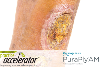
Biofilm can exacerbate a wound’s chronicity. Some estimate that biofilm is present in more than 80% of chronic wounds. Left untreated, biofilm will adhere to a wound surface and mature into a wound infection.5 Biofilm can become resistant at 48 to 96 hours after formation, inhibiting typical therapeutic wound healing modalities. Treating biofilm early on and repeatedly is necessary to keep biofilm at bay and promote continuous wound healing. Several modalities may be effective in treating biofilm.
Sharp debridement is a very effective way to remove biofilm and disrupt the wound bed by physically removing unhealthy tissue, senescent cells and biofilm. Antimicrobial dressings and cleansing agents may also kill or disrupt biofilm. When biofilm is left undisturbed in a wound, bacteria form and impede healing; dressings and cleansing agents that contain hypochlorous acid, polyhexamethylene biguanide (PHMB), silver, acetic acid, honey or iodine target biofilm effectively. Cellular and/or tissue-based products that contain antimicrobial components, such as silver or PHMB can be used to encourage closure while providing a sustained antimicrobial barrier to support bioburden management. Clinicians should verify that these modalities are within their scope of practice before using them.6

References
1. Basehore, B. M., Zito, P. M., & Wallace, H. A. (2020). Wound Healing Phases. Treasure Island, FL: StatPearls Publishing.
2. Territo, M. (2020, January). Monocyte Disorders. Retrieved from Merck Manuals: https://www.merckmanuals.com/home/blood-disorders/white-blood-cell-diso…
3. Alberts, B., Johnson, A., Lewis, J., Raff, M., Roberts, K. & Walter, P. (2002). Molecular Biology of the Cell. New York: Garland Science.
4. Cullen, B., Gibson, D., Harding, K., Legerstee, R. & Shultz, G. (2009). MMPs Made Easy. Wounds International, 1(1), 1-6.
5. Attinger, C. & Wolcott, R. (2012). Clinically Addressing Biofilm in Chronic Wounds. Advances in Wound Care, 1(3), 127-132. 6. Bjarnsholt, T., Eberlein, T., Malone, M. & Schultz, G. (2017). Management of Wound Biofilm Made Easy. Wounds International, 2(8), 1-6.
The views and opinions expressed in this content are solely those of the contributor, and do not represent the views of WoundSource, HMP Global, its affiliates, or subsidiary companies.
More from this Author
// fixed missing link variable.
// fixed missing link variable.
// fixed missing link variable.


