Lower Extremity Wounds: Is It Venous, Arterial, Neuropathic, or a Combination?
October 21, 2021
Keywords
Categories
Lower extremity wounds manifest in a multitude of ways, with numerous causative or trigger factors. These types of wounds are often costly to treat, are frequently refractory, and have a high risk for recurrence. A comprehensive assessment and an evidence-based treatment plan, along with ongoing patient education and routine follow-up, are essential components of an effective plan of care.1
Importance of Differential Assessment
As discussed in previous blogs, the first steps in effective wound management for any type of wound are to determine etiologic factors and to correct them before proceeding further. It is necessary to understand the cause before we can create and implement an effective plan of care. This is especially important to remember when differentiating among venous, arterial, and neuropathic wounds because often treatments for one wound type could be contraindicated or of no benefit when treating one of the other wound types.1
General Patient Assessment and History
Patient interview and physical assessment are key in formulating differential diagnoses when looking at venous, arterial, and neuropathic ulcers.
- Deep vein thrombosis, obesity, a sedentary lifestyle, and an impaired calf muscle pump are known risk factors for venous insufficiency.
- Coronary artery disease, hyperlipidemia, hypertension, diabetes, and tobacco use are known risk factors for arterial insufficiency.
- Long-standing diabetes and metabolic disorders are the most common known risk factors for neuropathy and neuropathic ulcers in the United States.1
Given that these wounds are often present in older adults with multiple, pre-existing comorbid conditions, it is imperative that the health care provider complete a thorough interview and patient history when determining wound etiology.
Assessing Comorbid Conditions
Certain health conditions may be present in conjunction with venous or arterial wounds. Some of these conditions may further complicate the treatment of lower extremity ulcers. Therefore, a thorough health history and acknowledgment of these conditions, along with involving the appropriate interprofessional team members, are key to establishing an effective plan of care. Venous ulcers may be present in the setting of heart failure, lymphedema, orthopedic procedures, and hypercoagulable states. Arterial ulcers can be associated with cardiovascular disease, cerebrovascular disease, and spinal cord injuries. Neuropathic ulcers can be present in patients with underlying arterial and renal disease. It is important to be aware of these conditions and adjust the plan of care accordingly.1
Pain
Venous ulcer pain is typically described as achy, heavy, and dull, involving the entire extremity. Pain is usually worse in the dependent position, and edema improves with elevation. Many patients report minimal pain in the morning if the legs have been elevated in bed throughout the night, and then pain worsens throughout the day.1 Arterial ulcer pain is usually described as cramping, intense, and throbbing, and it typically involves the lower leg and foot. Early in the disease, pain usually occurs with significant activity and is relieved with rest (intermittent claudication). In more advanced disease, pain is usually present when the legs are in a neutral or horizontal position (such as during sleep or lying down) and is relieved in the dependent position. As the disease progresses, pain typically becomes more constant.1 Neuropathic ulcer pain is usually described as a pins and needles, burning, stinging, or electric shock type of pain, and it is sometimes worse at night. Pain may be reduced by walking and often requires pharmaceutical management.1
Lower Extremity Assessment
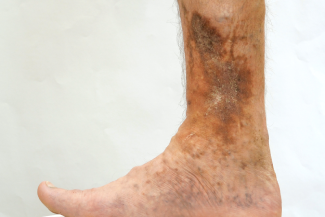
Both limbs should be assessed simultaneously, to compare the affected limb with the unaffected limb. Signs of venous compromise:
- Prominent ankle veins and varicosities
- Hemosiderin staining, usually to bilateral lower extremities
- Pitting edema, which usually extends ankle to knee
- Lipodermatosclerosis
- Venous dermatitis (erythema and/or pruritus from above the malleoli and below the calf muscle)1
Signs of arterial compromise:
- Thin, shiny skin
- Diminished or absent hair growth
- Rigid or thin nails
- Elevational pallor (legs paler when elevated)
- Dependent rubor (legs redder or pinker when in a dependent position)
- Coolness of the distal limb and foot
- Capillary refill time >3 seconds
- Diminished or absent pulses in the lower extremities
- Ankle brachial index <0.5, which may indicate critical limb ischemia
- Patients who keep their legs in the dependent position often may also have some noticeable dependent edema.1
Signs of neuropathy:
- Typically, a loss of protective sensation leading to failure to sense the monofilament at one or more sites when using the 10-g Semmes-Weinstein monofilament test1
Wound Assessment
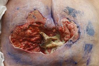
Wounds of different etiologies vary in their clinical features. Manifestations of venous wounds:
- These wounds are located on the lower extremity.
- They have moderate to heavy exudate, especially in the setting of edema, and the exudate may be clear or serosanguinous.
- Open areas are usually shallow and irregularly shaped, but wounds can also be full-thickness.
- The wound bed appearance is usually red or dark red, with or without slough.
- Slough and/or biofilm may be present because of high bacterial loads.
- There is typically not tunneling or undermining in these wounds.1
Manifestations of arterial wounds:
- These wounds are usually located on the toes, forefoot, or heels (areas most distal to the heart).
- These wounds can manifest as non-healing, traumatic injuries because of the lack of blood flow to the extremity to promote healing.
- Drainage is usually scant or absent in these wounds.
- Open areas are usually punched out or round in appearance, with well-defined borders.
- The wound bed appearance is usually pale or necrotic.1
Manifestations of neuropathic wounds:
- These wounds are usually located on the plantar aspect of the foot or metatarsal head of the great toe, dorsal toe, distal toe, or interdigital areas.
- Drainage is typically moderate to heavy unless the etiology is mixed.
- Wound beds are usually pink or red unless there is a mixed etiology or existing ischemia.1
Key Points
Patient history (including health history and medications) is critical when formulating a plan of care and determining wound etiology. Ulcer onset and history, along with pain onset and history (exacerbating and relieving factors), are important to understand as well. Additionally, wound location plays a major role in understanding etiology—venous ulcers are typically located between the ankle and the knee, and arterial ulcers are usually located more distally—toes or forefoot—most distal from the heart. Neuropathic ulcers are most commonly found on the plantar aspect of the foot.1 In future blogs, keep an eye out for differential assessment and management, clinical presentation of patients with mixed etiology wounds, and involvement of different disciplines for the most effective plan of care.
Reference
1. McNichol L, Ratliff C, Yates S. Differential assessment of lower extremity wounds. In: McNichol LL, Ratliff CR, Yates SS, eds. Wound, Ostomy and Continence Nurses Society Core Curriculum: Wound Management. 2nd edition. Philadelphia, PA: Wolters Kluwer; 2022:446-453.
About the Author
Holly is a board certified gerontological nurse and advanced practice wound, ostomy, and continence nurse coordinator at The Department of Veterans Affairs Medical Center in Cleveland, Ohio. She has a passion for education, teaching, and our veterans. Holly has been practicing in WOC nursing for approximately six years. She has much experience with the long-term care population and chronic wounds as well as pressure injuries, diabetic ulcers, venous and arterial wounds, surgical wounds, radiation dermatitis, and wounds requiring advanced wound therapy for healing. Holly enjoys teaching new nurses about wound care and, most importantly, pressure injury prevention. She enjoys working with each patient to come up with an individualized plan of care based on their needs and overall medical situation. She values the importance of taking an interprofessional approach with wound care and prevention overall, and involves each member of the health care team as much as possible. She also values the significance of the support of leadership within her facility and the overall impact of great teamwork for positive outcomes.
The views and opinions expressed in this content are solely those of the contributor, and do not represent the views of WoundSource, HMP Global, its affiliates, or subsidiary companies.
More from this Author
// fixed missing link variable.
// fixed missing link variable.
// fixed missing link variable.