Prevention of Heel Pressure Injuries: A Daunting Challenge
June 17, 2021
Keywords
Categories
Pressure injuries (PIs) are defined by the National Pressure Injury Advisory Panel as “localized damage to the skin and/or underlying soft tissue usually over a bony prominence or related to a medical or other device.” Pressure injuries may present as intact skin or as an open ulcer. These wound may be painful. Pressure injuries occur after exposure to prolonged pressure or as a result of pressure in combination with shear. Other factors may affect soft tissue tolerance, such as nutrition, perfusion, microclimate, the presence of comorbidities, and the condition of the soft tissue.1
Pressure Injury Etiology
PIs are a result of several interacting factors at play. Compression of soft tissue between two surfaces such as a bony prominence and a firm surface leads to a deformation injury of soft tissue cells that irreversibly damages the cell membrane and cytoskeleton (supporting structure of the cell). The pressure and shear forces result in inflammation, ischemia, and ultimately cell death if the pressure is not alleviated. Tissue injury from pressure, shear, and ischemia leads to ischemia-reperfusion injury over time. The effects of reperfusion worsen damage in the two to five days after circulation has returned. In addition, changes in the microclimate of the skin occur in response to the accumulation of moisture and heat, which increases the metabolic demand on cells and weakens the intercellular connections, thus reducing the tolerance of the tissues.
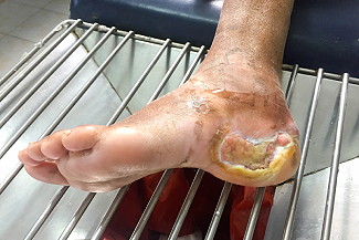
The heel is the second most common anatomical site for PI following the sacral area in all considered ages. Despite being so common, the pathophysiology of heel PIs (HPIs) is not yet completely understood. The angular anatomy of the calcaneus and its thin fat pad increase the risk of pressure injury development. In older adults, the dermal-epidermal junction is compromised, resulting in damage from friction forces. Other factors, such as reduced perfusion caused by microvascular and/or macrovascular disease, as well as impaired sensation from neuropathy or other forms of nerve damage, along with immobility, make for a perfect storm for HPI formation.
 Caution: Deep tissue injury
Prevention of Heel Pressure Injuries
The geometry, anatomy, and perfusion of the heel create challenges in preventing HPIs. International guidelines suggest that to redistribute pressure from the heel, the heel should be floated from the bed. Suspending or “floating” the heels clear off the support surface by elevating the lower leg or calf is demonstrated to be an effective strategy to relieve pressure, shear, and friction forces, thereby reducing the risk of HPIs. Unfortunately, floating the heel on pillows does not always work if the patient moves about in bed or the pillow collapses from the weight of the leg. In addition, floating the heels will always result in greater weight bearing in other anatomical areas and may shift the risk for injury to elsewhere in the body. When determining which mechanism to use to offload the heel, consider the following issues: How long will the legs be immobile, and how mobile is the patient? Is the patient agitated and moving a lot? Is there poor arterial flow? Are there sensory limitations such as paraplegia or neuropathy? Is the patient is ambulatory? Does the patient slide down in the bed, or do they need a bed extender? Considerations in Elevating the Heels2
- Ensure that a heel suspension device is not applied too tightly, or determine whether there is a risk of HPIs developing as a result of device application (medical device–related pressure injury).
- Inspect skin underneath the device at regular intervals.
- Avoid using water-filled gloves or intravenous fluid bags to elevate the heels because they place undue pressure on the Achilles tendon.
- Avoid pressure damage to the Achilles tendon by elevating the entire calf. Avoid popliteal vein compression and risk of deep vein thrombosis by positioning the knees in 5° to 10°. Consider the effect of the device on the skin microclimate. If the device significantly increases moisture and skin temperature, it may not be appropriate.
- Use a pressure redistribution support surface in conjunction with heel elevation.
- Keep in mind that if a patient is on a pressure redistribution surface, they still need to have the heels floated.
Specific Devices for Heel Elevation
Heel suspension boots designed from egg-crate foam to suspend the foot on an elevated pad within a protected boot space that extend up the lower leg can be effective in reducing the incidence of HPIs. Heel suspension boots designed from polyurethane foam have been trialed in older adults. It appears that there is no significant difference between the different models of heel suspension boot for efficacy in preventing HPIs. Both can be effective as long as they are maintained in proper position.
Foam cushions can also be of value. A foam block cushion can be employed to support the entire lower leg, thereby floating the heels, used with a second foam block that supports the feet to prevent foot drop. Standard pillows can also be as effective as a heel suspension boot in preventing HPIs; however, a standard pillow may be unreliable in maintaining the heels in an elevated position for extended periods of time, especially in more mobile individuals or in those with dementia, agitation, or leg spasms. Foam dressings have been used in selective settings such as intensive care units and perioperative environments. Application of a multi-layer polyurethane foam dressing with a silicone border can reduce the interface pressure between the heel and a standard viscoelastic hospital mattress.
Conclusion
HPIs tend to occur more often in immobile patients. The risk factors for these injuries stem from the anatomy of the calcaneus, impairments in blood flow to the foot, and neuropathic disease. There are many clinical considerations in the prevention of HPI. Although the use of preventive interventions is critical, it is imperative to continue to closely monitor the patient for the development of HPIs. In more compromised patients, it is still possible to develop HPIs even with optimal care. Close surveillance and accurate documentation of HPIs and progression are critical.
References
1. Edsberg LE, Black JM, Goldberg M, McNichol L, Moore L, Sieggreen M. Revised National Pressure Ulcer Advisory Panel pressure injury staging system: revised pressure injury staging system: Revised pressure injury staging system. J Wound Ostomy Continence Nurs. 2016;43(6):585-597.
2. Haesler E. Evidence summary: pressure injuries: preventing heel pressure injuries with positioning. Wound Pract Res. 2017;25(4):212-214.
Resources
Black J, Santamaria N, Gefen A, Brindle T, Fletcher J, Alves P. Prevention and management of pressure injury to the heel . Wounds International.com. 2018:43-49.
Rivolo M, Dionisi S, Olivari D, et al. Heel pressure injuries: consensus-based recommendations for assessment and management. Adv Wound Care (New Rochelle). 2020;9(6):332-347.
VanGilder C, Lachenbruch C, Algrim-Boyle C, Meyer S. The International Pressure Ulcer PrevalenceTM survey: 2006-2015: a 10-year pressure injury prevalence and demographic trend analysis by care setting. J Wound Ostomy Continence Nurs. 2017;44(1):20-28.
The views and opinions expressed in this content are solely those of the contributor, and do not represent the views of WoundSource, HMP Global, its affiliates, or subsidiary companies.
More from this Author
// fixed missing link variable.
// fixed missing link variable.
// fixed missing link variable.