What Are the Advantages of Multilayer Compression Bandaging for Chronic Venous Insufficiency, Lymphedema, and Phlebolymphedema?
Keywords
Categories
Background
As lymphedema and wound care therapists at Penn Therapy & Fitness, we often see scenarios like those in the cases described in our abstract presented as a poster at the recent Symposium on Advanced Wound Care. Conditions such as lymphedema, chronic venous insufficiency, and phlebolymphedema that manifest in patients with wounds can lead to catastrophic and life-threatening skin breakdown (degradation). Complications of infections associated with lymphedema include cellulitis, lymphangitis, lymphadenitis, and ulcerations where protein-rich fluid provides a perfect medium for microbial growth. Challenges can also develop when there are:
- Nutritional challenges
- Poor hygiene
- Poor skin care management
- Decreased movement and activity
Considerations for Lymphedema and Phlebolymphedema Treatment
Lymphedema is an accumulation of protein-enriched fluid in the interstitial space resulting from an imbalance of net ultrafiltration at the capillary level. Phlebolymphedema is caused by insufficiency of the venous or lymphatic system (or both), in combination with possible systemic contributors, leading to accumulation of protein-rich fluid in the interstitial space.
A patient should consider contacting a clinician if they experience the following:
- Swelling of part or all of the arm or leg, including fingers or toes
- A feeling of heaviness or tightness
- Restricted range of motion
- Recurring infections
- Hardening and thickening of the skin (fibrosis)
Venous Ulcer Cause and Characteristics
Cause
Incompetent venous valves create lower extremity edema secondary to increased venous hypertension resulting in ulceration.
Location
Venous ulcers develop in the gaiter area, extending just above the malleolus to below the knee. They tend to occur on both lateral and medial aspects of the leg.
Characteristics
- Dorsal foot sparing
- Round open sore in the skin, with the outer surface possibly raised and thick
- Weeping or blistering
- Hemosiderin staining (skin discoloration)
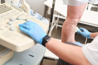
Evaluating Patients With Phlebolymphedema
When evaluating patients with phlebolymphedema, the examination includes an assessment of the patient’s history and review of systems, including comorbidities that would otherwise indicate a history of cellulitic, cardiac, or vascular conditions, as well as the initial onset of disease.
Physical assessment of patients with phlebolymphedema include:
- Capillary refill test
- Palpation and assessment of dorsalis pedis and posterior tibial pulses
- Stemmer’s sign (usually negative at the toes if caused more by venous insufficiency than by lymphatic mechanical insufficiency)
- Assessment of tissue texture
Objective measurements in patients with phlebolymphedema include:
- Circumferential girth measurements
- Fluid displacement
- Perimeter
Other aspects of the evaluation include the following: length, width, and depth measurements; level of slough or eschar; odor; assessment of periwound; and level of wound drainage. It is also important to rule out deep vein thrombosis (DVT) from vascular disorders, especially if the patient has a history of DVT.
Our Treatment Approach
The lymphatic system plays a role in vascular wound healing, as does effective compression bandaging. As shown in our poster presentation, our patients received complete decongestive therapy (CDT), which is the gold standard of care for lymphedema. We also selected a novel multilayer compression bandaging approach—foam-based wound care dressings and Fuzzy Whale compression technology (EdemaWear). This therapy helped improve venous return and microcirculation, release vasoactive mediators, and decrease peripheral congestion and tissue remodeling to prevent ulcer recurrence.
Handout for Patients
If you notice any skin or fluid changes, talk to your doctor about seeing a Certified Lymphedema Therapist (CLT) to address the swelling, regardless of how minimal or severe the edema appears to be. In fact, if the swelling and wounds are addressed sooner, especially in the earlier stage, you may require less intensive treatment or less time spent in the intensive phase of CDT. If your swelling is getting you down and the wounds are draining and leaking all over the place, talk to your doctor about your symptoms. A CLT and wound care specialist may be what you need to reduce the edema and heal the wounds so you can start enjoying life.
About the Authors
Tia Gray PTA,, CLT, CWT is a Physical Therapist Assistant at Good Shepard Penn Partners. Tia is also a Certified Lymphedema and Wound Care Specialist. Tia received her Associates in Physical Therapy from Harcum College.
Dr. Donald Thomas PT, DPT, CLT-LANA, CWT is a Physical Therapist at Good Shepard Penn Partners. Dr. Thomas is also a certified Lymphedema and wound care specialist. He received his Doctorate in physical therapy from Widener University.
The views and opinions expressed in this content are solely those of the contributor, and do not represent the views of WoundSource, HMP Global, its affiliates, or subsidiary companies.

