Acute Treatment of Extravasation Injuries in Neonates: Polymeric Membrane Dressings
February 1, 2023
Keywords
Categories
Introduction
Hospitalized patients of all ages are at risk for iatrogenic injuries. Peripheral intravenous extravasation and infiltration (PIVIE) injuries are one of the most common types of hospital-acquired injury, with the potential to cause long-term disfigurement or loss of function. While the administration of fluids and medications through intravenous catheters is a common practice in the hospital, it is not without significant risk if fluids leak from the vasculature into surrounding tissues. Depending on the type and amount of fluid that enters the tissue, varying degrees of tissue damage may result. The pathophysiology of this tissue damage may also vary.1
PIVIEs in Neonates
Pediatric patients, neonates in particular, have unique characteristics that place them at a higher risk for PIVIE-related injuries, including the following1-4:
- The immature skin, subcutaneous tissues, and vasculature in neonates are much thinner and more fragile than in older children and adults.
- Frequent assessment of vascular access device sites may be more challenging in pediatric patients. Devices may be covered with layers of tape or elastic wraps to prevent dislodgement.
- Neonates are unable to effectively communicate specific pain, which may be an early indicator that a vascular access device is malfunctioning.
The occurrence rates of PIVIE-related injuries in pediatric patients are largely unknown. Literature reviews report rates ranging from 2% to 70% of neonates affected by extravasation injuries.1-4 Pediatric hospitals in the United States are only just beginning to formally track, monitor, and implement prevention programs for PIVIEs. Included in the bundle of PIVIE prevention measures from the Solutions for Patient Safety Network is hourly assessment of the site for early recognition of complications, as well as education of the patient’s family to identify and report concerns regarding vascular access sites by observing and touching the site and comparing it to the other extremity.5 Early signs of infiltration or extravasation may include the following:
- Edema
- Erythema
- Temperature change
- Pain at the site of a peripheral intravenous (PIV) device
Leakage of blood or fluid from the insertion site may be observed. If extravasations are not detected early, more severe signs of skin and tissue damage may result. These include pallor and blanching, areas of very dark discoloration, bullae formation, or extreme edema with tight, shiny skin. In my experience, PIVIEs that show acute discoloration of the skin (ie, very dark purple/maroon or very light white/gray) most often progress to a full-thickness wound with eschar.
Treatment Options for PIVIE
Treatment methods for extravasation injuries vary widely, and there have been no formal studies to evaluate the effectiveness of treatments reported in literature. Among the most common recommendations for acute treatment of PIVIE include the following:
- Aspiration of any fluid remaining in the access catheter
- Removal of the device
- Elevation of the extremity
In addition to the above, an antidote should be administered as soon as possible, typically within 12 hours of discovery. Hyaluronidase may be administered subcutaneously to make local tissues more permeable for dispersion of the extravasate. Phentolamine may be selected as an antidote when vasoconstrictive medications have extravasated. Guidelines for the use of these antidotes in neonates are unclear and often left up to individual prescribers.
The use of hot or cold compresses is debated, with little evidence to support either practice. More invasive treatment practices may include saline flush-outs or perforation of the skin with a needle or scalpel to allow drainage of excess fluid.1-4 Discussion of treatment practices for extravasation injuries in neonates is even more sparse and mostly limited to case studies. A general consensus about all extravasation injuries is that treatment should begin immediately to reduce pain, edema, tissue damage, and subsequent wound formation.1 Numerous recommendations also suggest consulting a specialist to guide treatment of extravasation injuries and their sequelae. Topical applications and wound dressings are not considered a part of the acute management phase of extravasation treatment, but possibly should be.
A Novel Approach: Polymeric Membrane Dressings
Clinicians should consider the unique properties of the various wound dressings available to meet each patient’s individual needs. With limited recommendations available for the acute management of extravasation injuries, it is beneficial to have a wound care expert to guide and oversee treatment. Topical treatment options may be limited during the acute phase, while skin discoloration is evolving and demarcating into a more defined area of tissue damage.
Polymeric membrane dressings represent a unique category of wound dressings that have been shown to reduce tissue edema and inflammation, even in closed injuries like sprains and bruises. Polymeric membrane dressings are also the only drug-free dressing shown to reduce pain.6 These innovative properties set polymeric membrane dressings apart from standard foam dressings and led me to explore their benefit in the acute management of extravasation injuries. This period of time is critical since the tissue damage is evolving, but as care providers, we often just have to “watch and wait” for a more defined wound to develop. Consider the following case study as an example of the use of polymeric membrane dressings to treat extravasation injuries.
Case Study
Our WOCN team was consulted to evaluate an extravasation injury on the wrist of a 4-week-old baby. The extravasate contained epinephrine and vasopressin infused through a peripheral intravenous access device. An antidote was not administered. On my initial assessment, an area of very dark purple discoloration (approximately 4cm x 3cm) was noted on the ventral aspect of the left wrist, see image A of Figure 1. The skin was intact. In my previous experience, I have seen numerous injuries with similar presentations, and all have progressed to full-thickness wounds with eschar.
Typically, the area of discoloration is monitored until tissue damage is well-demarcated, and then moist wound healing is initiated with various topical products. In this case, a single layer of polymeric membrane dressing was applied around the wrist to cover the visible area of tissue damage. The dressing was left in place for several days at a time and changed as needed when it was visibly soiled or at nurses' discretion. The nursing care order stated to change the dressing when soiled or saturated, but since there was no open wound under the dressing, it did not become soiled and could be left in place for extended periods. The dressing was secured with a small piece of silicone tape so that the dressing was in contact with the skin, but there was no pressure around the wrist.
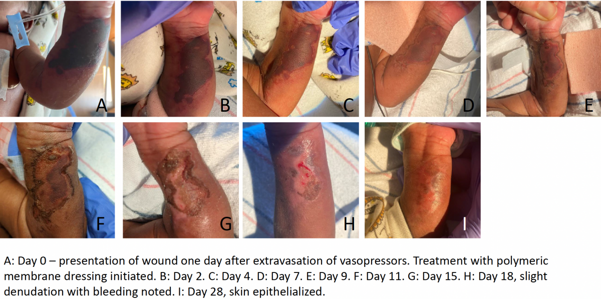
For 4 weeks, the wound was reassessed 3 times weekly by our team. We observed a steady reduction in the size and intensity of skin discoloration. Very slight, superficial peeling of the skin was noted around the edges of the skin discoloration as it decreased in size. On day 18, I noted a small spot of denudation with scant bleeding (see Image H of Figure 1). Treatment with the polymeric membrane dressing was continued, and complete healing was observed on day 28. Due to the patient’s age, his pain level could not be accurately assessed, but no analgesic medications were given during any wound dressing changes.
Conclusion
This case study exhibits the successful outcome of a neonatal patient with a severe extravasation injury who was successfully treated with polymeric membrane dressings, beginning in the acute phase. Based on my previous experience with similar patients and the similar initial appearance of PIVIE, this patient had a significantly shorter healing time. This patient did not experience progression to eschar or a full-thickness wound, which I have previously observed in similar cases. The use of polymeric membrane dressings presents no risk of harm to the patient. Additionally, the dressings' ability to stay in place for several days makes it a cost-effective treatment option. The unique properties of polymeric membrane dressings to reduce tissue edema, inflammation, and pain should be further investigated as a treatment option for extravasation injuries.
References
- Hackenberg RK, Kabir K, Muller A, Heydweiller A, Burger C, Welle K. Extravasation injuries on the limbs in neonates and children – development of a treatment algorithm. Dtsch Arztebl Int. 2021;118:547-54.
- Odom B, Lowe L, Yates C. Peripheral Infiltration and Extravasation Injury Methodology: A Retrospective Study. J Infus Nurs. 2018;41(4):247-252.
- Dufficy M, Takashima M, Cunningham J, et al. Extravasation injury management for neonates and children: A systematic review and aggregated case series. J Hosp Med. 2022;17:832-842.
- Corbett M, Marshall D, Harden M, Oddie S, Phillips R, McGuire W. Treatment of extravasation injuries in infants and young children: a scoping review and survey. Health Technol Assess. 2018;22(46):1-112.
- Children’s Hospitals’ Solutions for Patient Safety Network (SPS Network). Agency for Healthcare Research and Quality; 2017. Accessed February 1, 2023. https://www.ahrq.gov/workingforquality/priorities-in-action/sps-network…
- Dechant ED. Considerations for Skin and Wound Care in Pediatric Patients. Phys Med Rehabil Clin N Am. 2022;33(4):759-771.
About the Author
Elizabeth Day Dechant, BSN, RN, CWOCN, CFCN is a Certified Wound Ostomy Continence Nurse at Children’s of Alabama, where she provides wound treatment recommendations and wound management for both inpatients and outpatients with acute and chronic wounds. She provides staff education on skin and wound care, ostomy care, and pressure injury prevention. Elizabeth works diligently with the hospital’s Pressure Injury Prevention Team to track and reduce hospital-acquired pressure injuries.
The views and opinions expressed in this content are solely those of the contributor, and do not represent the views of WoundSource, HMP Global, its affiliates, or subsidiary companies.
More from this Author
// fixed missing link variable.
 // fixed missing link variable.
// fixed missing link variable.
// fixed missing link variable.