Evaluation of Surgical Wounds
Practice Accelerator
September 22, 2023
September 22, 2023
Keywords
Categories
Introduction
Successful care of surgical wounds begins with a comprehensive assessment of both the wound and the patient, including a clear understanding of the type and class of surgical wound. Psychosocial factors affecting healing and well-being, are essential to consider as well.1
Definitions and Classifications
Surgical wounds can be categorized through the following:
- Incisional or excisional
- Wound healing type
- Infection risk
Surgical wound classification enables wound care professionals to tailor a care plan to each patient’s risk of complications.2
Wound healing type describes the method of wound closure as follows2-4:
|
Healing Type |
Definition |
Comments |
|
Primary intention |
Closed wound (all layers closed) with approximated wound edges |
Most common surgical wound type Rapid healing Minimal scarring and tissue loss |
|
Secondary intention |
Wound with deep layers. Closed and superficial layers left open to heal from the bottom up because of infection or an inability to approximate wound edges |
Slow healing Higher infection risk More extensive scarring
|
|
Tertiary intention |
Delayed primary closure, with the wound left open for several days initially |
Exudate drainage Control of contamination Preparation for further procedures |
The Centers for Disease Control and Prevention (CDC) classifies surgical wounds by the degree of wound contaminants, as shown here2-4:
|
Class |
Definition |
|
Class I/ Clean |
Operative wounds (usually skin, eyes, or vascular) with no signs of infection or inflammation No internal organ involvement Surgical site infection risk < 2% |
|
Class II/Clean-contaminated |
Operative wounds with no outward evidence of infection and no break in sterile technique Internal organ involvement Surgical site infection risk <10% |
|
Class III/Contaminated |
Open, fresh, accidental wounds involving internal organs or operative wounds with a break in sterile technique, gastrointestinal tract spillage, or incisions with acute, nonpurulent inflammation Also open traumatic wounds >12-24 hours old Surgical site infection risk <13%-30% |
|
Class IV/Dirty |
Wounds infected during operation, with visceral perforation or acute purulent inflammation Also delayed presentation of traumatic wounds with contamination and devitalized tissue already present Surgical site infection risk almost 40% |
Assessment Protocol
The following assessment protocol is partly adapted from the South West Regional Wound Care Program (London, Ontario, Canada)1:
- Obtain a complete medical history, including medications, nutrition, wound history and pain, wound healing factors, and the patient’s goals and involvement in care.
- Perform a general physical examination, including a current pain assessment.
- Perform a psychosocial assessment, including the patient’s wound comprehension, financial concerns and support, the patient’s environment, and the patient and family’s functional, cognitive, and emotional ability to manage care.
- Note incision anatomic location and length, closure method (sutures, staples, glue, pins, or other external fixation6), closure status, hemorrhage, inflammation, condition of wound bed and periwound, and surgical drains.
- Complete a validated wound assessment tool (eg, the National Pressure Injury Advisory Panel PUSH [Pressure Ulcer Scale of Healing] Tool 3.05) and repeat at least weekly to monitor wound healing and care.
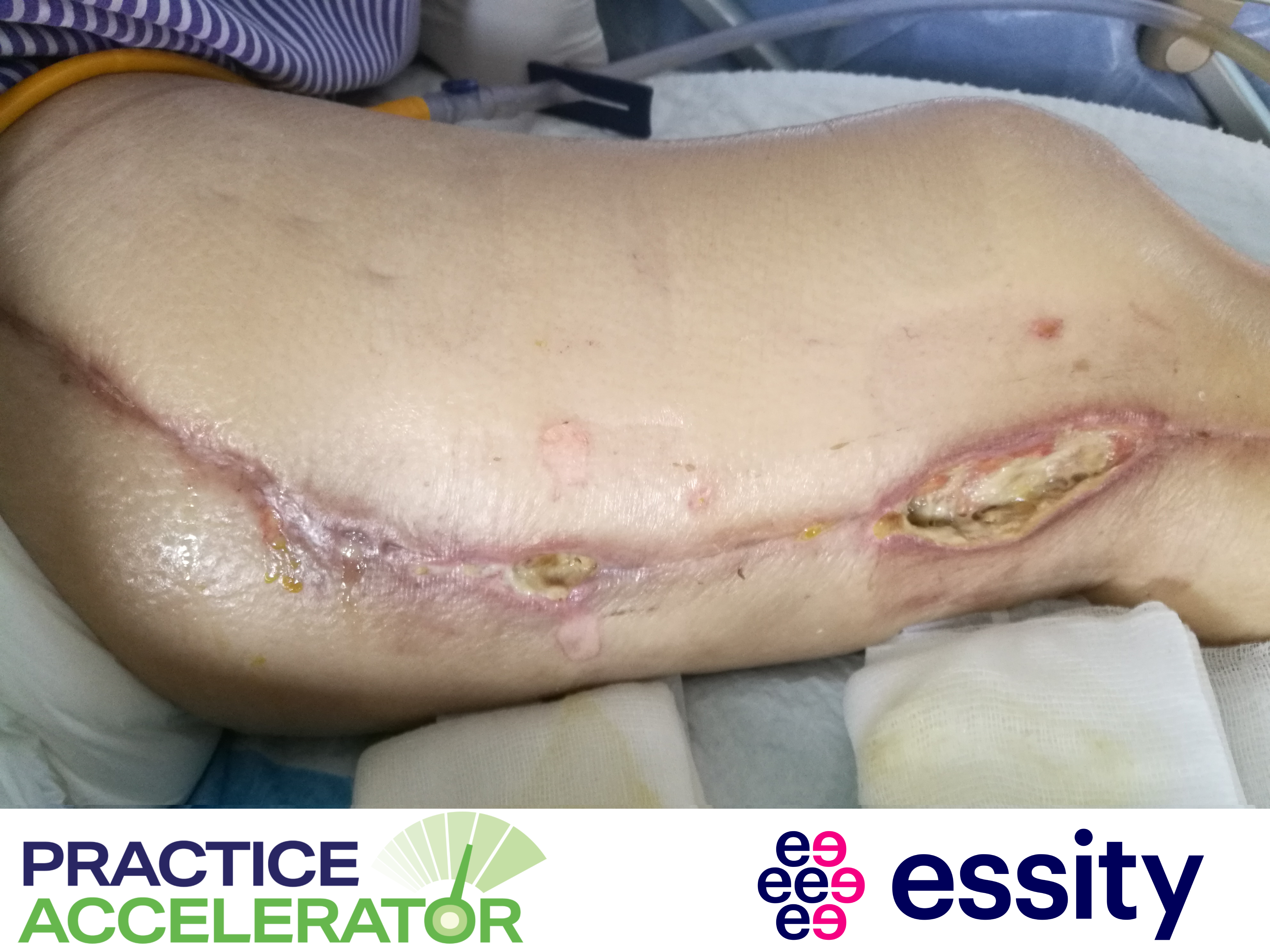
- Assess the wound for increased bioburden, including hemopurulent or seropurulent wound drainage 48 hours postoperatively, incision site abscess or breakdown, or spontaneous dehiscence.
- Assess wound moisture balance and dressing appropriateness.
- If the surgical wound is on the lower leg, assess the leg for edema, lymphedema, lipidema, vascular disease, and circulation.
- If a patient with diabetes and/or neuropathy has a surgical foot wound, obtain the patient’s treatment history and diabetic complications. Assess the patient’s feet and toenails for deformity, range of motion, neuropathy, and infection. Also assess gait, footwear, and edema, lymphedema, lipidema, vascular disease, and circulation.
- Assess and monitor any sutures, staples, and glue sites,7 as well as external fixation devices,6 and make sure an order is in place for removal. Timing of suture (nonabsorbable) or staple removal varies from 3 to 21 days.3
- Determine wound healability.
- Document all findings meticulously.
Monitoring for Surgical Site Infection
Monitoring can prevent or minimize surgical site infections. Patients with symptoms of infection (surgical site pulling, persistent fatigue and malaise, warmth with pain, and foul odor) should contact their health care provider or surgeon immediately.8 The risk for complications is multifactorial and includes contamination, mechanism of injury contamination, systemic involvement, and presurgical conditions.2 For these reasons, knowledge of definitions, classifications, and assessment protocols for is essential to preventing and managing these challenging wounds effectively.
References
- South West Regional Wound Care Program. Surgical wounds assessment guide. 2020. Accessed September 2, 2023. https://www.swrwoundcareprogram.ca/
- Surgical wounds 101. Practice Accelerator. WoundSource. 2018. Accessed August 31, 2023. https://www.woundsource.com/blog/surgical-wounds-101
- American College of Surgeons. Wound Home Skills Kit: Surgical Wounds. 2018. Accessed August 31, 2023. https://www.facs.org/for-patients/home-skills-for-patients/wound-manage…
- Herman TF Bordoni B. Wound classification. In: StatPearls [Internet]; 2022. Accessed August 31, 2023. https://www.ncbi.nlm.nih.gov/books/NBK554456/
- Swezey L. Wound assessment tools. WoundSource. 2015. Accessed August 31, 2023. https://www.woundsource.com/blog/wound-assessment-tools-basic-introduct…
- Clinical guidelines (nursing): Pin site care for the child with an external fixator. Royal Children’s Hospital Melbourne. Accessed September 3, 2023. https://www.rch.org.au/rchcpg/hospital_clinical_guideline_index/Pin_sit…
- Azmat CE, Council M. Wound closure techniques. StatPearls [Internet]. 2023. Accessed September 2, 2023. https://pubmed.ncbi.nlm.nih.gov/29262163/
- Assessment of surgical wound infections. Practice Accelerator. WoundSource. 2019. Accessed September 2, 2023. https://www.woundsource.com/blog/assessment-surgical-wound-infections
The views and opinions expressed in this content are solely those of the contributor, and do not represent the views of WoundSource, HMP Global, its affiliates, or subsidiary companies.
More from this Author
// fixed missing link variable.
// fixed missing link variable.
// fixed missing link variable.


