Diagnosis and Pathophysiology of Venous Leg Ulcers
Practice Accelerator
January 24, 2020
January 24, 2020
Keywords
Categories
The most common type of chronic lower extremity wound is the venous ulcer, affecting 1% to 3% of the U.S. population.1,2 Chronic venous ulcers significantly impact quality of life and are a financial burden for both the patient and the health care system. In the United States, 10% to 35% of adults have chronic venous insufficiency, and 4% of adults 65 years old or older have venous ulcers.3 Identifying signs of venous disease early on while implementing surgical intervention, if warranted, can increase healing outcomes and decrease the recurrence of venous ulcers. Treatment of venous ulcers can include exercise, leg elevation, dressings, advanced wound care such as cellular and tissue-based products, compression therapy, medications, venous ablation, and surgical intervention.4
Pathophysiology of Venous Ulcers
Early identification of the risks and signs of vascular disease can help prevent new and recurring ulcers, as well as infection. The primary underlying mechanism of venous ulcer formation is venous reflux, which is increased venous pressure, also known as venous hypertension. Venous hypertension results from incompetent valves or obstruction in the macrocirculation. Blood pools in an area of the extremity, and an ulcer develops as a consequence. Exudate may fluctuate from minimal to heavy, depending on edema management and/or infection.4
Venous Ulcer Identification
- Location: gaiter area (i.e., area extending just above the ankle to below the knee; may occur on both lateral and or medial aspects)5
- Shape: irregular wound margins
- Depth: shallow
- Tissue: may contain granulation, slough, eschar, and/or fibrin
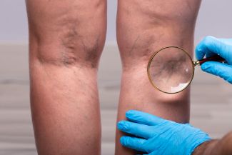
- Varicose veins
- Symptoms of leg heaviness and pain6
- Pruritus6
- Edema
- Venous dermatitis
- Telangiectasias
- Hemosiderin staining
- Corona phlebectatica
- Atrophie blanche
- Lipodermatosclerosis
- Deformity of leg: often described as looking like an upside-down champagne bottle on the lower extremity
Predisposing Comorbidities
There are many comorbidities that, alone or together, contribute to the risk of developing venous disease.4
- Age older than 55 years
- Family history
- Higher body mass index
- History of pulmonary embolism
- History of superficial or deep vein thrombosis
- Lower extremity skeletal or joint disease
- Higher number of pregnancies
- Physical inactivity
- History of ulcers
- Damaged venous system (intravenous drug users, recipients of intravenous therapy)
- Parental history of ankle ulcers
- Severe lipodermatosclerosis
- Venous reflux in deep veins
Diagnostic Testing
The diagnosis of venous ulcers is made based on ulcer anatomical location, characteristics, and morphology. Validation of venous disease requires laboratory studies as indicated, functional assessment of the venous system, and diagnostic tests. The "gold standard" for diagnosing venous disease is venography, but because of its cost, its associated morbidity, and the availability of non-invasive tests, it is performed infrequently. The venous duplex test is the method most often used to diagnose venous disease.7
Appropriate Antibiotic Use
Venous ulcers that are chronic or have prolonged healing are at higher risk for bacterial colonization, biofilm formation, and infection. Systemic antibiotics are used frequently to treat venous ulcers, but these drugs should be used only when there are signs and symptoms of infection.8,9
Conclusion
Identifying risks and signs of vascular disease early on can help with prevention of new and recurring ulcers, as well as infection. When venous ulcers become chronic, refer to appropriate specialists, such as a vascular surgeon and wound care specialist, to work together in developing the best treatment plan.
References
1. Ruckley CV, Evans CJ, Allan PL, et al. Chronic venous insufficiency: clinical and duplex correlations. The Edinburgh Vein Study of venous disorders in the general population. J Vasc Surg. 2002; 36(3):520-525.
2. Lal BK. Venous ulcers of the lower extremity: definition, epidemiology, and economic and social burdens. Semin Vasc Surg. 2015;28(1):3-5.
3. Rabe E, Guex JJ, Puskas A, Scuderi A, Fernandez Quesada F; VCP Coordinators. Epidemiology of chronic venous disorders in geographically diverse populations: results from the Vein Consult Program. Int Angiol. 2012;31(2):105-115.
4. Bonkemeyer Millan S, Gan R, Townsend PE. Venous ulcers: diagnosis and treatment. Am Fam Physician. 2019;100(5):298-305.
5. Chi YW, Raffetto JD. Venous leg ulceration pathophysiology and evidence-based treatment. Vasc Med. 2015;20(2):168-181.
6. Vivas A, Lev-Tov H, Kirsner RS. Venous leg ulcers (Japanese version). Ann Intern Med. 2016;165(3):JITC17-JITC32.
7. Min RJ, Khilnani NM, Golia P. Duplex ultrasound evaluation of lower extremity venous insufficiency. J Vasc Interv Radiol. 2003;14(10):1233-1241.
8. Bryant R, Nix D. Acute and Chronic Wounds: Current Management Concepts. 3rd ed. St. Louis, MO: Elsevier Mosby; 2006:162.
9. Bowler PG, Duerden BI, Armstrong DG. Wound microbiology and associated approaches to wound management. Clin Microbiol Rev. 2001;14(2):244-269.
The views and opinions expressed in this content are solely those of the contributor, and do not represent the views of WoundSource, HMP Global, its affiliates, or subsidiary companies.
More from this Author
// fixed missing link variable.
// fixed missing link variable.
// fixed missing link variable.


